. 2020 Jul 23;2(4):e200312. doi: 10.1148/ryct.2020200312

Figure 3b:

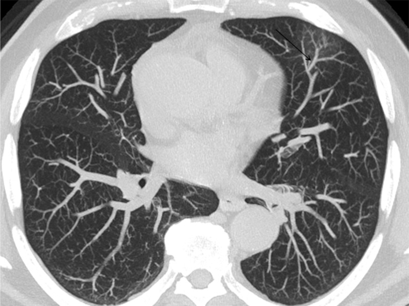
A 74-year-old man arriving at the emergency department with fever. (a) The CT scan documented a focal ground-glass opacity in the superior segment of the lingula (circle), classified as an “indeterminate” pattern. Notably, there was a vascular enlargement in the pulmonary artery branch afferent to the lesion, as (b) the maximum intensity projection better demonstrated (arrow). The real-time reverse-transcription polymerase chain reaction test was positive for severe acute respiratory syndrome coronavirus 2 infection.

A 74-year-old man arriving at the emergency department with fever. (a) The CT scan documented a focal ground-glass opacity in the superior segment of the lingula (circle), classified as an “indeterminate” pattern. Notably, there was a vascular enlargement in the pulmonary artery branch afferent to the lesion, as (b) the maximum intensity projection better demonstrated (arrow). The real-time reverse-transcription polymerase chain reaction test was positive for severe acute respiratory syndrome coronavirus 2 infection.