Figure 6.

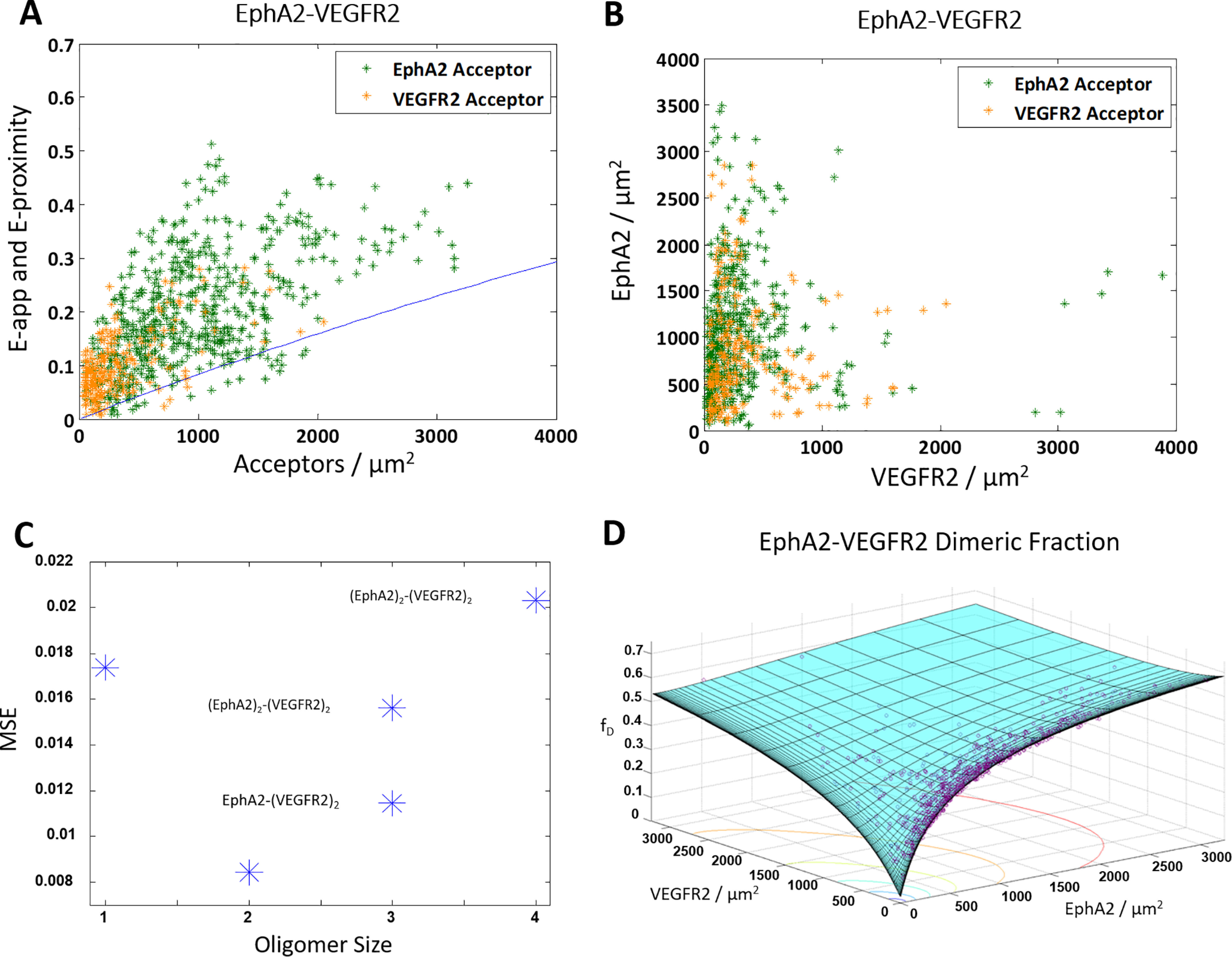
The truncated ECTM EPHA2 and ECTM VEGFR2 form a heterodimer. HEK 293T cells were transiently co-transfected with versions of EPHA2 and VEGFR2 where the IC domain has been replaced with a fluorophore: either MT for the donor or eYFP for the acceptor. The cells were transfected with varying ratios of EPHA2 and VEGFR2, with a total of 0.5–3 μg of EPHA2 DNA and 3–6 μg of VEGFR2 DNA. A, apparent FRET efficiency versus acceptor concentration. Shown are 856 data points (592 for VEGFR2–MT and EPHA2–eYFP (green) and 264 for EPHA2–MT and VEGFR2–eYFP (orange)). The solid blue line is the monomer proximity FRET (60, 61). B, EPHA2 concentration versus VEGFR2 concentration. C, an MSE plot, showing that the heterodimer model gives the best fit. D, dimeric fraction as a function of VEGFR2 and EPHA2. The purple symbols are the experimentally determined dimeric fractions, and the solid cyan surface is the best-fit surface for the heterodimer model.
