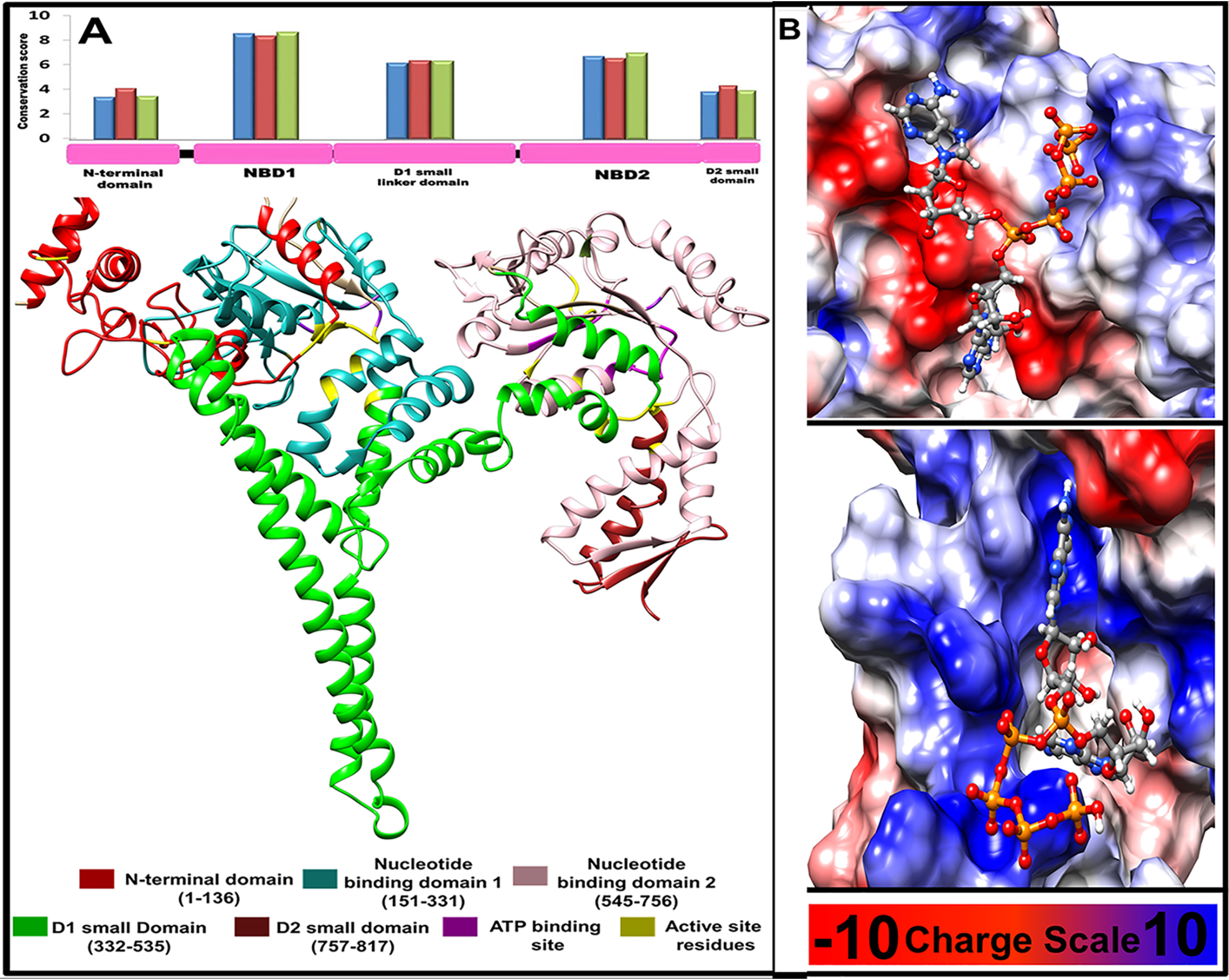
Figure 6.

Structural features of L. donovani HSP78 protein. A, 2D and 3D domain organizations of L. donovani HSP78 protein and their relative conservation. Blue, red, and green bars show conservation of individual domain calculated using the representative sequences from all phyla, Euglenozoa, and Ascomycota, respectively. B, binding mode and probable interactions of the selected inhibitor Ap5A, which shows significant binding potential for NBD1 and NBD2 shown in upper and lower panel, respectively.
