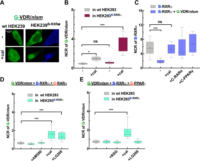
Figure 6.
EGFP-VDR/nlsm as NR1 in competition with RARα and PPARγ: both RARα and PPARγ dominate over VDR/nlsm in competing for dimerization with RXRα. A, representative confocal images of G-VDR/nlsm transiently transfected into WT HEK293 (left) or HEK293B-RXRα cells (right) in the absence (top) or presence (bottom) of specific NR1 agonists (10−7 M cal). Scale bar, 10 μm. B, nuclear-to-cytoplasmic fluorescence intensity/pixel ratios (NCR) of G-VDR/nlsm. C, NCR values of B-RXRα. D, NCR of G-VDR/nlsm in HEK293B-RXRα cotransfected with C-RARα were assessed in the absence or presence of agonists (10−7 m AM580, 10−7 m cal, or 10−7 m LG268). E, changes in the distribution of G-VDR/nlsm in HEK293B-RXRα cotransfected with C-PPARγ were assessed in the absence or presence of agonists (10−6 m RSG, 10−7 m cal, or 10−7 m LG268). Box-and-whiskers plots represent 10th, 25th, 50th, 75th, and 90th percentiles; +, mean value. G, EGFP; B, TagBFP; C, mCherry. *, p < 0.05; ***, p < 0.0001; ns, not significant.

