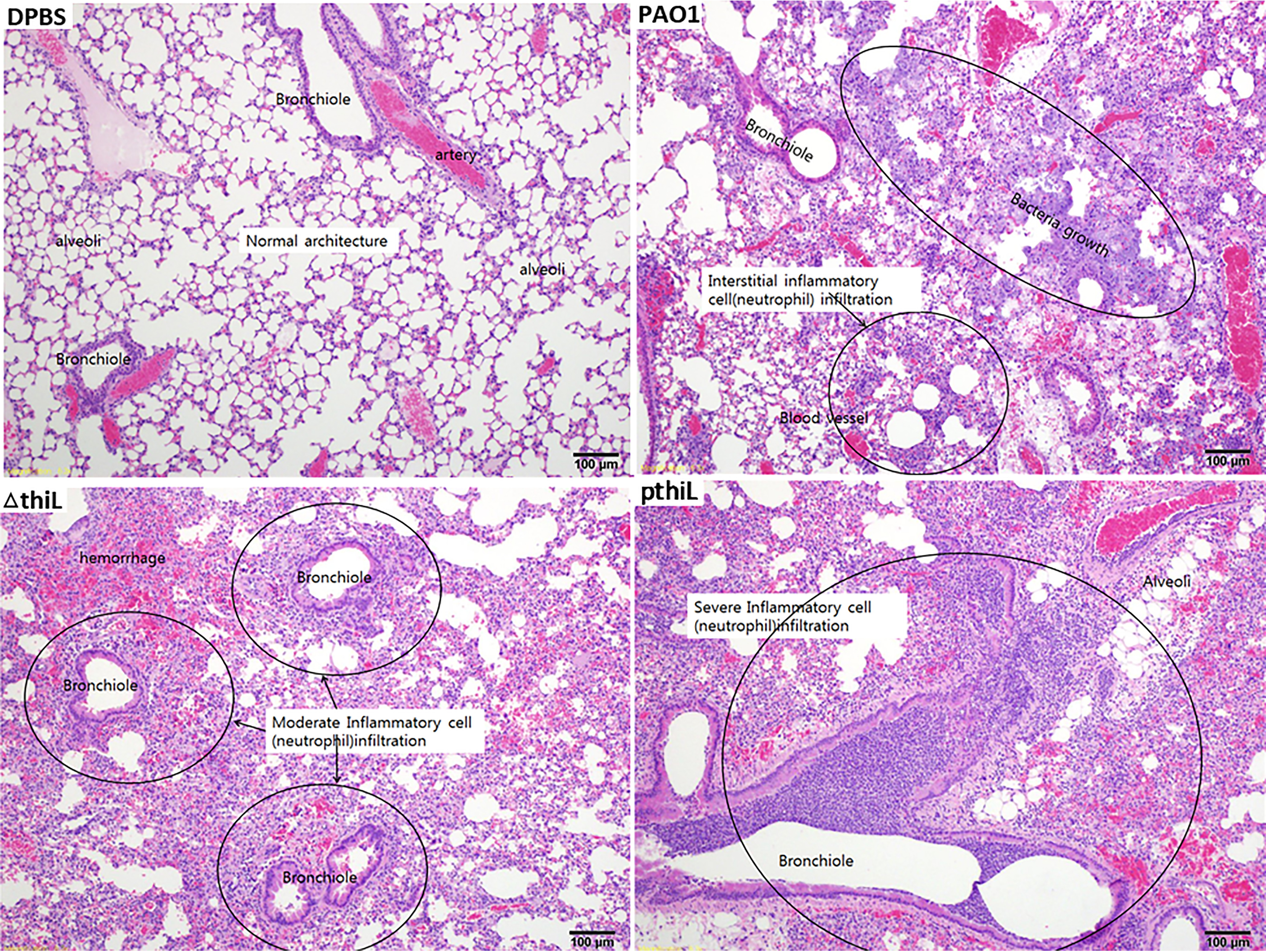
Figure 4.

Neutrophil infiltration of the left lobe of lungs in C57BL/6 mice infected with PAO1, ΔthiL, or pthiL. Intranasal infections were applied to 6-week-old mice with DPBS, PAO1, ΔthiL, or pthiL. H&E staining showed moderate to severe neutrophil infiltration (marked in circles) in bronchioles and blood vessels of the lungs after 20 h of infection. Magnification, ×100. Representative images of triplicate experimental groups are shown.
