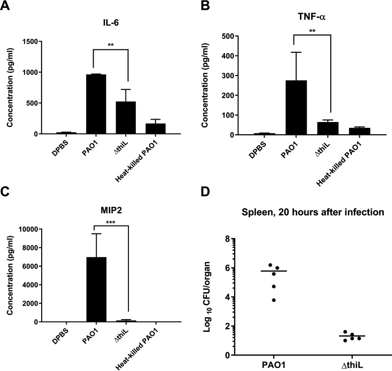
Figure 6.
Proinflammatory cytokine (IL-6, TNFα, and MIP-2) levels in the blood of mice 20 h after infection with PAO1, ΔthiL, or heat-killed PAO1 and bacterial loads (CFU/organ) in mouse spleen 20 h after infection with PAO1 or ΔthiL. A–C, ELISAs for IL-6 (A), TNFα (B), and MIP-2 (C) in mouse plasma indicated significantly lower cytokine levels in ΔthiL groups, compared with PAO1. **, p < 0.01; ***, p < 0.001. D, CFU analysis showed 104-fold lower bacterial loads in the spleens of mice infected with ΔthiL, compared with PAO1 (n = 5 for each experimental group, repeated three times).

