
Figure 2. Structural features of Mango-III.
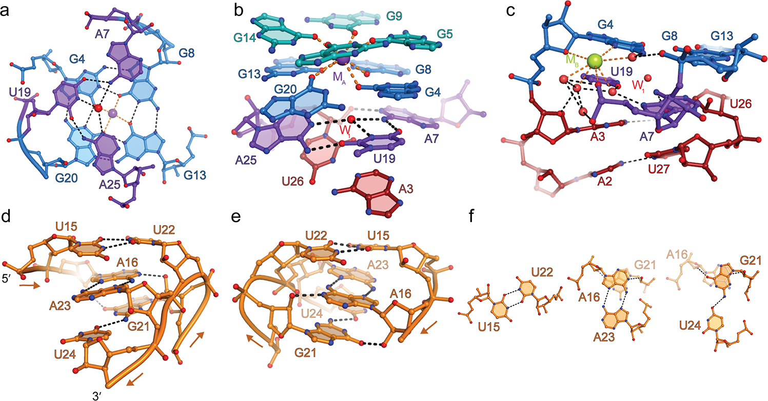
(a) Hydrogen bonding interactions of the A7•U19•A25 base triple (purple) and the G-quadruplex T1(blue). Red and purple spheres, water molecule at the center of the triple (W1), and K+ at the center of the G-quadruplex (MA), respectively. (b) The base triple stacks under the G-quadruplex and above the duplex P1. (c) Hydrated metal ion modeled as Na+ (green sphere) bridging the coaxial junction. (d) View of P2 from the direction of the G21-U24 strand. Arrows denote 5’ to 3’ chain direction. (e) 180° rotation along the vertical axis. (f) The three non-canonical base pairs of P2.