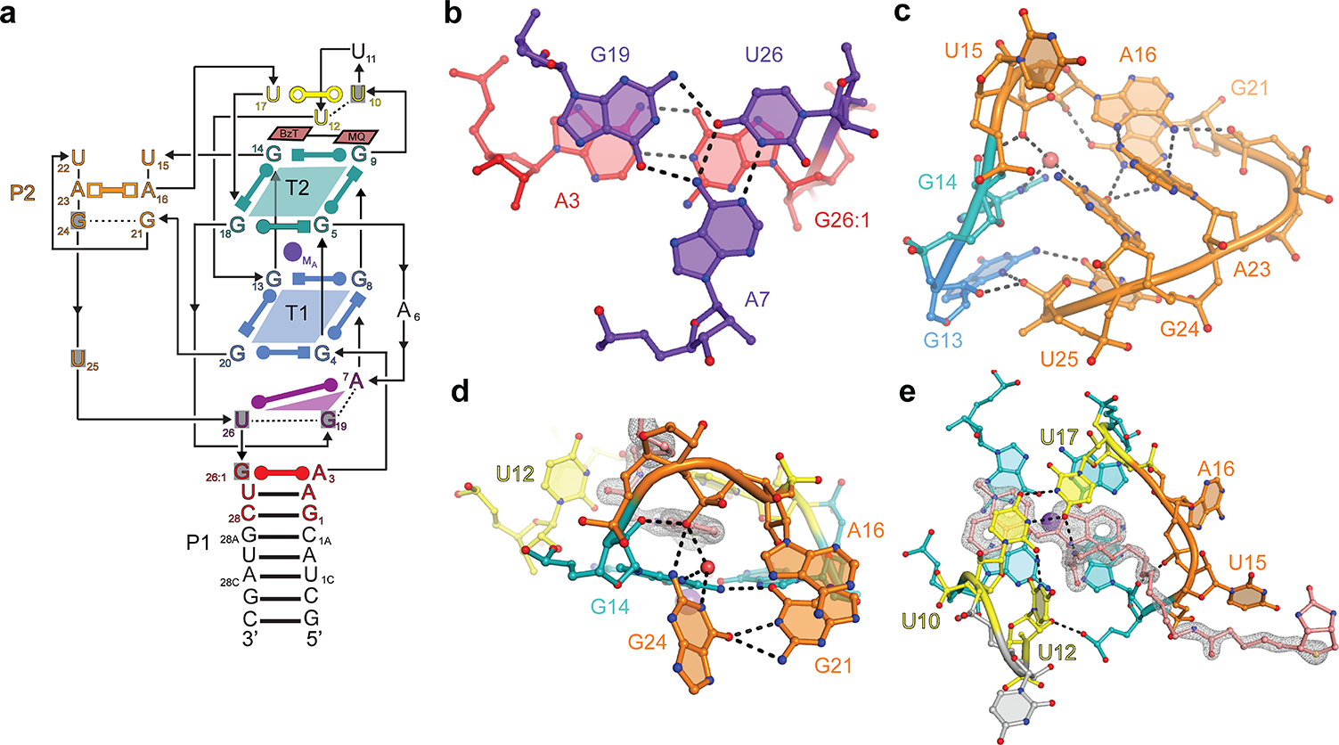
Figure 5. Structure of iMango-III.

(a) Schematic secondary structure of iMango-III. Residues that differ from the Mango-III sequence are shaded gray. (b) Structure of the rearranged junctional base triple. (c) Structure of the rearranged P2. (d) Interaction of the G21•G24 base pair with T2 of the G-quadruplex. (e) TO-1-Biotin recognition by iMango-III. Gray mesh, |Fo| - |Fc| electron density prior to building the fluorophore (2.5 s.d.)
