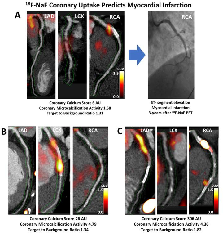
Figure 2. Case examples of 18F-sodium fluoride positron emission tomography in patients with established coronary artery disease and myocardial infarction during follow-up.
Hybrid CT angiography and 18F-NaF positron emission tomography of coronary arteries in: (A) a 56-year-old male who demonstrated increased 18F-NaF uptake in the RCA at baseline and presented with an inferior ST-segment elevation myocardial infarction and occlusion of the RCA during follow-up; (B) a 52-year-old male who demonstrated increased 18F-NaF uptake in the LCx at baseline and presented with a lateral non-ST-segment elevation myocardial infarction during follow-up; (C) a 60-year-old female who showed increased 18F-NaF uptake in the proximal RCA and presented with an inferior non-ST-segment elevation myocardial infarction during follow-up. LAD–left anterior descending, LCx–left circumflex, RCA–right coronary artery.

