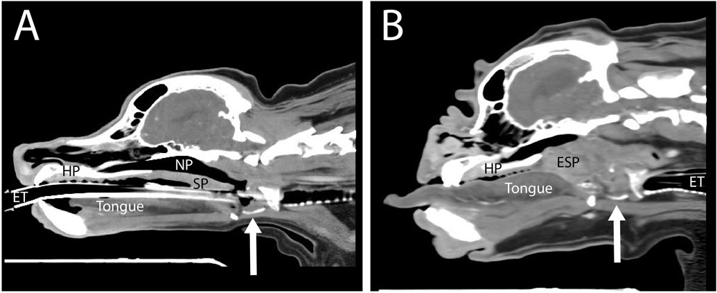
Fig. 4.
Airway soft tissue configurations. Computed tomography images of (A) mesaticephalic (cocker spaniel) and (B) brachycephalic (bulldog) dogs, at a sagittal plane slightly off midline, capturing the laryngeal cartilages at a similar level through the cricoid cartilage (arrow indicates the ventral cut section of the thyroid cartilage). In (B) note the following differences compared with (A): the thickened and elongated soft palate (ESP), the lack of patency of the nasopharyngeal (NP) airway to the level of the larynx, macroglossia and narrowed nasal airway. HP, hard palate; SP, soft palate; ET, endotracheal tube.

