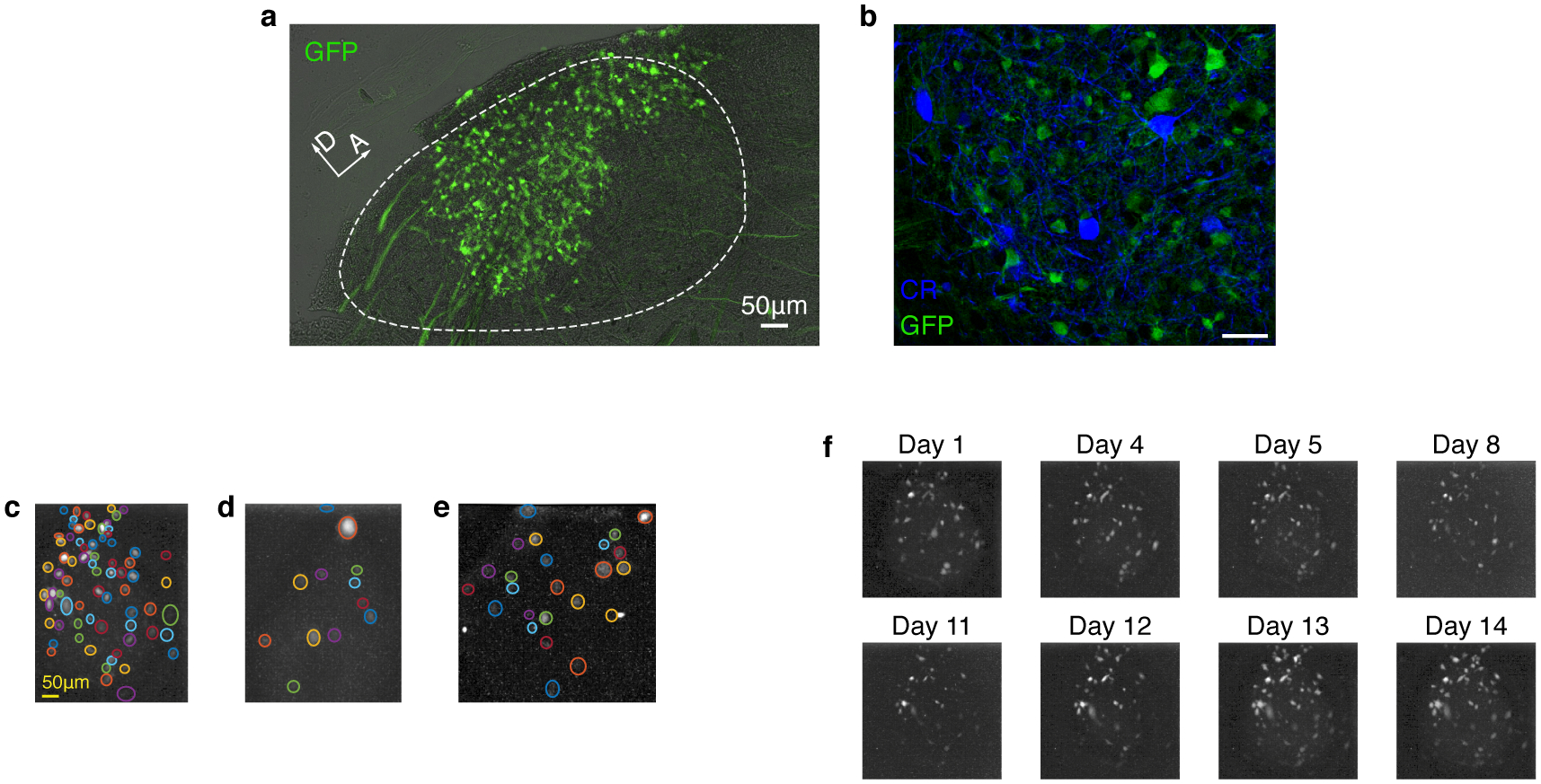
Extended Data Fig. 4 |. Calcium indicator is expressed exclusively in HVC excitatory neurons and imaged in annotated regions of interest (ROIs).

a. Sagittal slice of HVC showing GCaMP expressing projection neurons (Experiment repeated in 5 birds with similar results). b. We observed no overlap between transduced GCaMP6f-expressing neurons, and neurons stained for the inhibitory neurons markers calretinin, calbindin, and parvalbumin (CR stain shown, staining experiment repeated 6 times for each marker with similar results). c-e. Example of daily ROI annotation in 3 birds. Colored circles mark different ROIs, manually annotated on maximum fluorescence projection images an exemplary day (see methods). Panel are for birds 1–3. f. Maximum fluorescence images (methods, from bird 1) revealing the fluorescence sources including sparsely active cells in the imaging window across multiple days.
