
Figure 1 |.
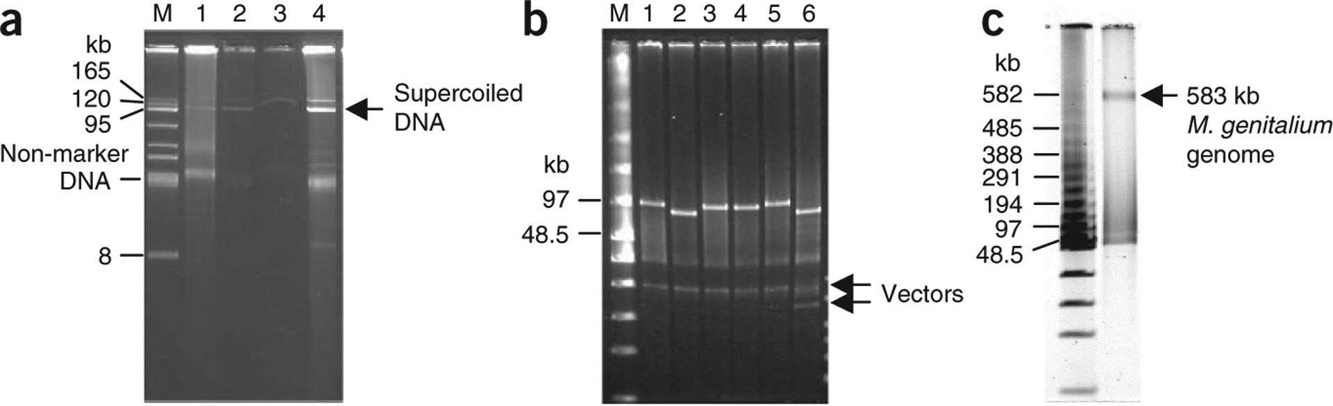
Purification of circular YAC from 400 ml of yeast culture. (a) Electrophoresis analysis of a 100-kb intermediate YAC of M. mycoides19, YAC 2a-100, recovered during purification. DNA was separated on a 1% agarose/1× TAE electrophoresis gel and stained as described in EQUIPMENT SETUP. M, BAC-tracker supercoiled DNA ladder; lane 1, 20 μl of DNA sample from Step 15 (before adding exonuclease); lane 2, 20 μl of DNA sample from Step 16 (after 35 min incubation); lane 3, 20 μl of the eluate (Step 21 before DNA precipitation); and lane 4, 1 μl of DNA sample from Step 25 (DNA pellet was dissolved in 150 μl of TE buffer). (b) NotI restriction enzyme digestion analysis of six YACs. A 0.75 μl sample of each YAC DNA was digested with NotI, which releases the insert DNA of YACs, and analyzed by FIGE. FIGE analysis was performed as described in EQUIPMENT SETUP. M, low-range PFG marker; lane 1, YAC 301–400 (~100 kb); lane 2, YAC 2a-100 (~96 kb); lane 3, YAC 101–200 (~100 kb); lane 4, YAC 401–500 (~100 kb); lane 5, YAC 501–600 (~100 kb); and lane 6, YAC 811a-900 (~88 kb). (c) NotI digestion analysis of ~600 kb YAC containing a synthetic M. genitalium genome17,18. A 0.75-μl DNA sample (from a total volume of 150 μl) was digested with NotI, which releases the M. genitalium genome (~580 kb) from the vector, and analyzed in a 1% (wt/vol) agarose gel by FIGE. The parameters were essentially as described in EQUIPMENT SETUP, with the following modifications: forward 90 V, initial switch 5.0 s, final switch 30 sec, with linear ramp.