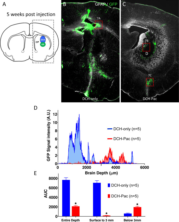
Fig 5. The DCH-paclitaxel system substantively ablates locally residing hGBM cells but does not prevent the migration of tumor cells within brain parenchyma.
A. Schematic of mouse forebrain showing the location of hGBM and DCH injections in the caudate putamen (CP). Box of dashed lines delineates the area quantitatively analyzed for the presence of GFP labeled cells in 5-week post injection tissue and presented in D and E. B,C. Survey immunofluorescent images of staining for GFP (hGBM cells) and GFAP show the location of hGBM cells in forebrain at 5 weeks post injection. B. Mouse that received hGBM cells and DCH-only. Note the high density of GFP-positive hGBM cells immediately above and below the injection site. C. Mouse that received hGBM cells and DCH-paclitaxel. Note the essential absence of GFP-positive hGBM cells immediately around the persisting DCH-paclitaxel depot (D). Note also the presence of hGBM cells that have migrated away from the injection site (box shown at higher magnification in 7C). D. Graph of quantification of GFP signal from hGBM cells. GFP staining intensity was measured as a function of brain depth in serial linear units in the boxed area shown in A. As expected, animals receiving DCH-only (blue) exhibited a high intensity of GFP signal in and immediately above the injection region (0.5 to 3 mm). In contrast, animals receiving DCH-paclitaxel (DCH-Pac, red) exhibited little or no GFP signal in this area, but did exhibit substantive signal at deeper levels (around 4mm). E. Bar graphs quantifying area under the Curve (AUC) for GFP staining intensity. Over the entire depth of the brain, DCH-paclitaxel treated animals (DCH-Pac, red) exhibited an over 72% reduction in hGBM-derived GFP signal compared with DCH-only (p<0.001). In the area immediately around the hGBM and DCH injections (surface to 3 mm), DCH-paclitaxel treated animals exhibited an over 97% reduction in hGBM-derived GFP signal (p<0.001). In contrast, in the deeper areas away from DCH depots (around 4 mm), DCH-paclitaxel treated animals exhibited a 65% greater hGBM-derived GFP signal (p<0.001). n = 5 per group for all measures.

