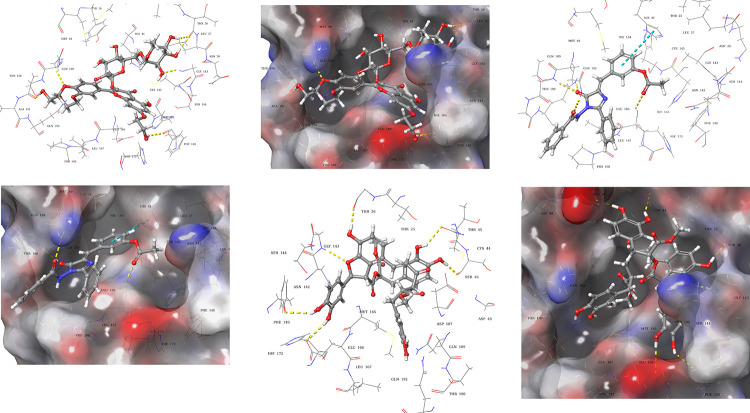
Fig 5. Docked poses and molecular interactions of compound 1, 2, and 3 in binding site of 6Y2F.
3D-Ligand interaction and 3D molecular surface diagrams of compounds 1 (a) and (b), compound 2 (c) and (d) compound 3 (e) and (f), showing the docked pose in the binding site of 2019-nCOV 3CL hydrolase (Mpro). The hydrogen bonds, and π-π interaction are represented as yellow and blue dotted lines.

