
FIGURE 5.
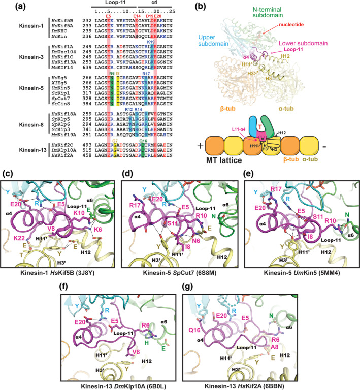
Comparison of the L11‐α4 region. (a) Sequence alignment of the L11‐α4 segments for selected kinesin family members. (b) Location of L11‐α4 within the motor domain and its interface with tubulin. (c–g) The conformation of L11‐α4 in each kinesin structure (magenta) and its contacts with α‐tubulin (yellow) are shown. Residue numbers in the kinesins have been changed to correspond to the columns in the sequence alignment for simplicity. PDB IDs are shown in parentheses