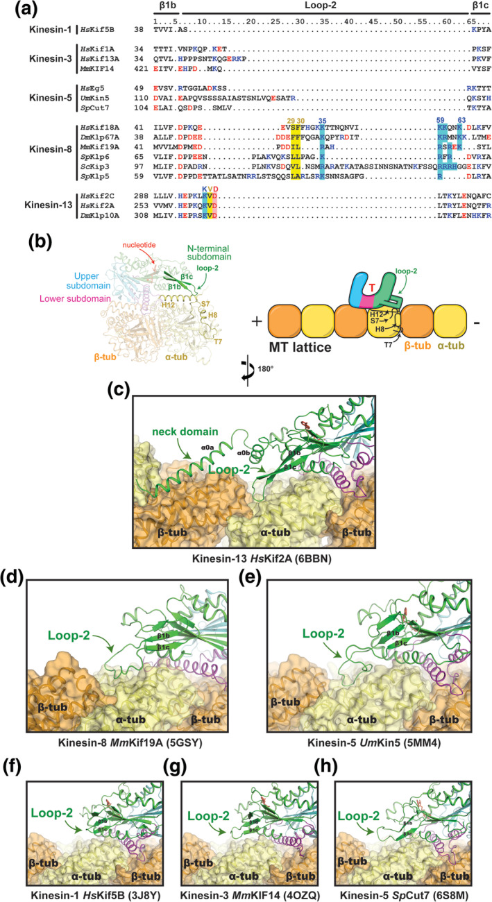
FIGURE 6.

Comparison of loop‐2 structures and MT interactions. (a) Sequence alignment of the loop‐2 region for selected kinesin family members. (b) Location of loop‐2 within the motor domain and its interface with tubulin. (c–h) The conformation of loop‐2 in each kinesin structure (green) and its contacts with tubulin (orange and yellow) are shown. PDB IDs are shown in parentheses. (g) The coordinates for the MmKIF14 motor domain (4OZQ) were docked onto the MT‐bound structure of apo kinesin‐1 (3J8X)
