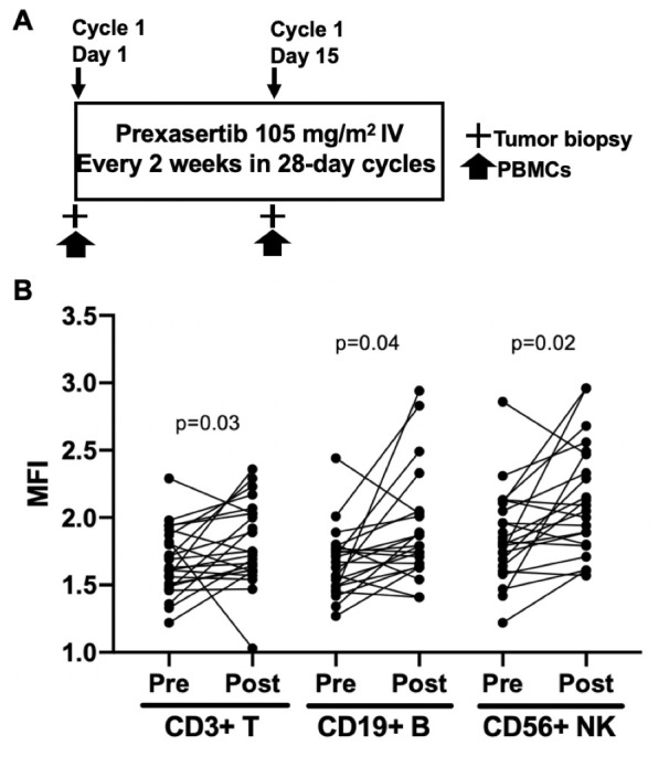
Figure 1.

Study schema and increased γ-H2AX, possibly caused by CHK1i. (A) Prexasertib was administered intravenously at 105 mg/m2 every 2 weeks in 28-day cycles until disease progression or unacceptable toxicities. A mandatory baseline tumor biopsy was performed before C1D1 treatment, and an optional second biopsy was performed 6 to 24 hours after treatment on C1D15. PBMCs for flow cytometry were collected at C1D1 and 6 to 24 hours after treatment on C1D15. (B) Flow cytometric analysis showing increased MFI of γH2AX in PBMC isolated from patients before (pre) and after a 2-week treatment with prexasertib (post). Cells were first gated on CD3 (T-cells), CD19 (B cells), or CD56 (NK cells) prior to plotting for γH2AX. C1D1, cycle 1 day 1; C1D15, cycle 1 day 15; MFI, mean fluorescence intensity; NK, natural killer; PBMCs, peripheral blood mononuclear cells.
