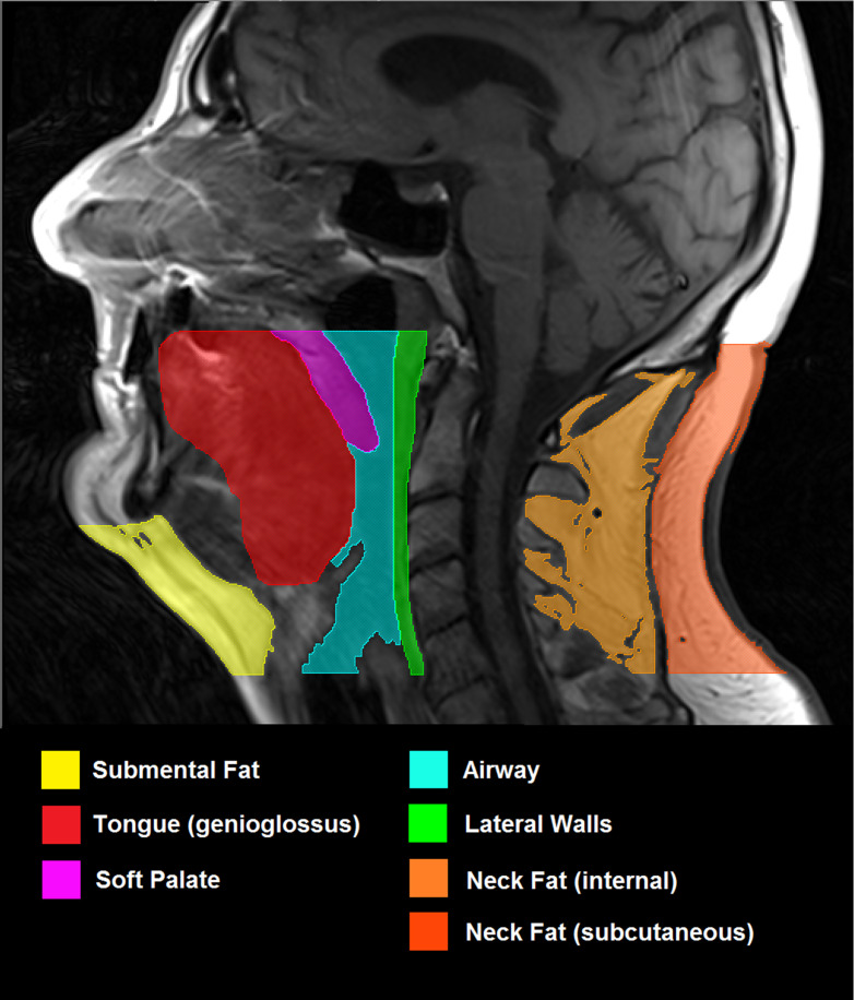
Figure 2.
MRI (T1-weighted, spin echo, 4 mm slice thickness) showing the mid-sagittal slice of head and neck. Region of interest includes all tissues inferior to hard palate and superior to vocal cords. The following structures to be included in analysis have been highlighted: submental fat, defined as all fat anterior to hyoid and inferior to mandible; tongue (genioglossus); soft palate; airway; lateral parapharyngeal walls; internal neck fat; subcutaneous neck fat.

