
Figure 2.
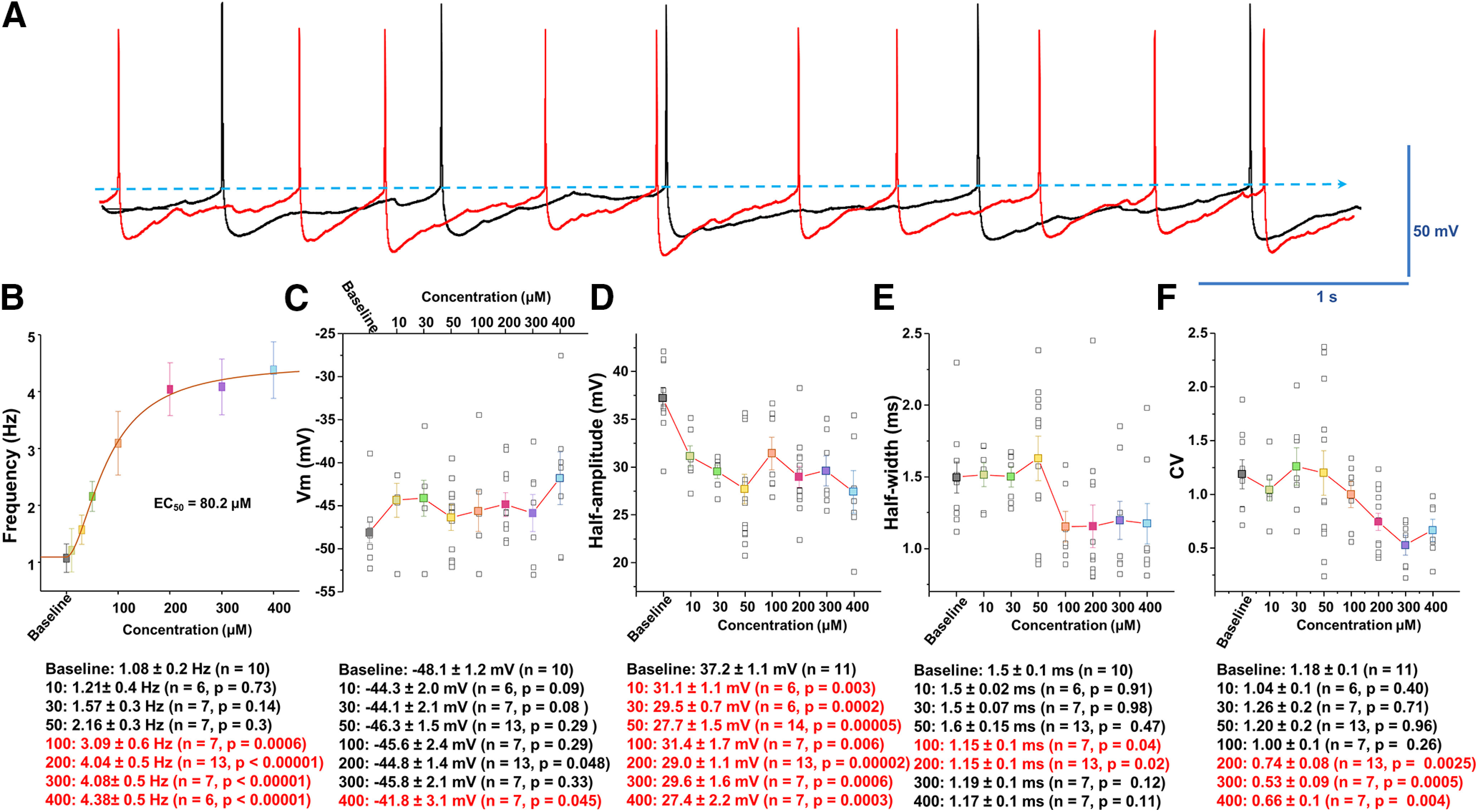
Analysis of spontaneous firing activity and properties of AP following manganese exposure. A, Representative AP trace before (baseline, black trace) and after bath application of 100 μm Mn2+ (red trace). B, Concentration-response relationships of the spontaneous firing frequency of dopamine neurons following Mn2+ exposure. An EC50 of 80.2 μm was obtained by fitting the concentration-dependent curve using a Hill equation. C, Mn2+ did not significantly change membrane potential. D, Mn2+ suppressed the amplitude of AP (as measured as the half-amplitude) at all concentrations examined (baseline: 37.2 ± 1.1 mV; 10 μm manganese: 31.1 ± 1.1 mV; t(15) = 3.6, p = 0.03, two-tailed Student's t test; n = 11 vs 6). E, Mn2+ did not change the half-width of AP at 10-50 μm, but it significantly narrowed the half-width at 100 μm (baseline: 1.5 ± 0.1 ms; 100 μm manganese: 1.15 ± 0.1 ms; t(15) = 2.2, p = 0.04, two-tailed Student's t test; n = 10 vs 7). F, Mn2+ concentration-dependently decreased the CV of interspike intervals (baseline: 1.2 ± 0.1; 200 μm manganese: 0.7 ± 0.1; t(22) = 3.4, p = 0.003, two-tailed Student's t test; n = 11 vs 13).