
Figure 4.
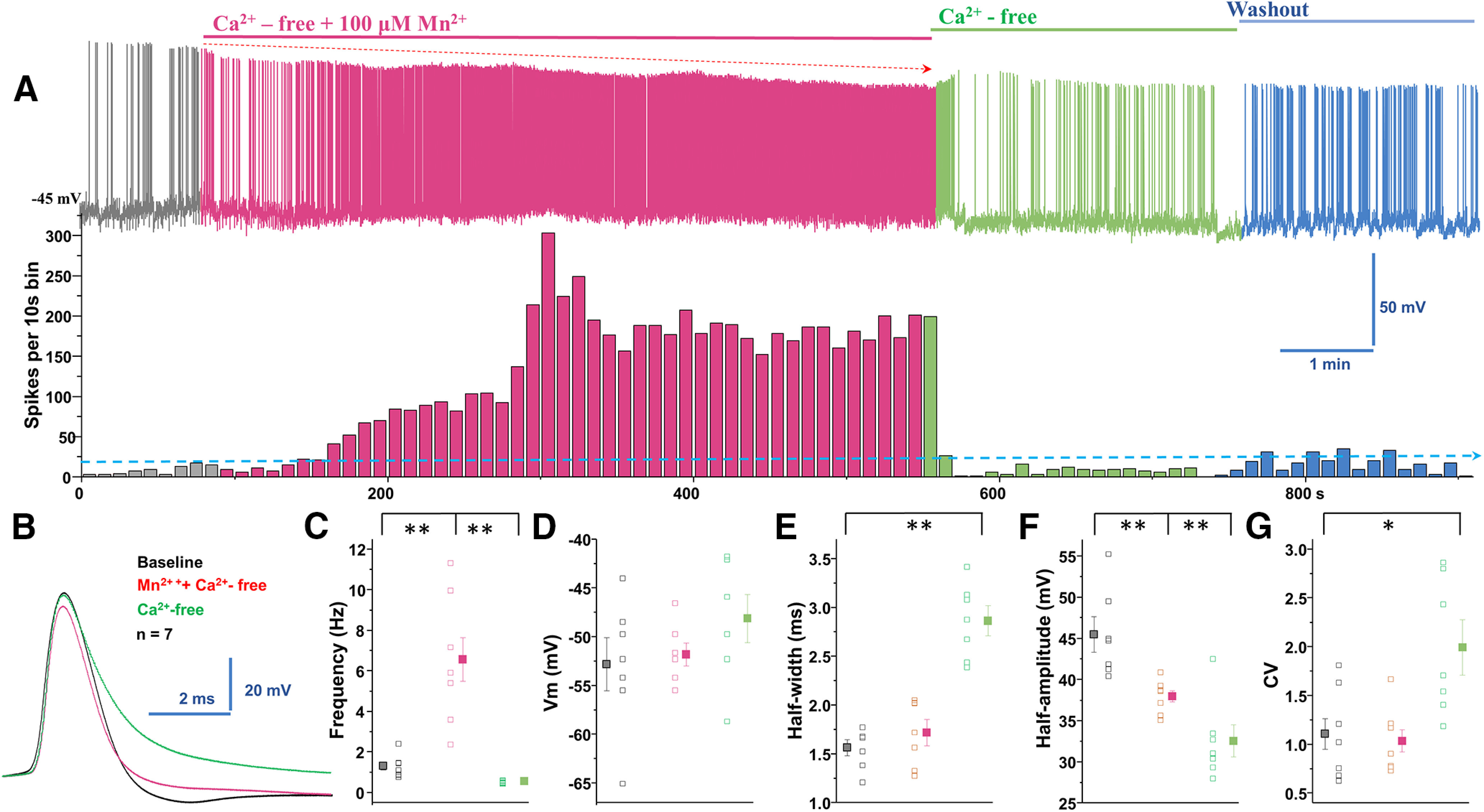
Manganese stimulation of firing activity of dopamine neurons is not dependent on extracellular Ca2+. A, Top, Representative spontaneous spike activities of a neuron exposed to Mn2+ in a Ca2+-free extracellular solution. Middle, Histogram of firing frequency obtained from the above trace showing Mn2+ increased firing frequency. B, Superimposed representative single AP traces shown in A, baseline (black trace), Mn2+ + Ca2+-free solution (red trace), and Ca2+-free solution only (green trace). C, In Ca2+-free solution, Mn2+ increased spontaneous firing rate. Mn2+ washout in Ca2+-free conditions revealed a reduction of firing rate (baseline: 1.3 ± 0.2 Hz vs manganese in Ca2+-free: 6.5 ± 1.1 Hz; F(2,18) = 20.9, p = 0.000002, one-way ANOVA followed by Tukey's test; n = 7/group). D, Mn2+ and Ca2+-free extracellular solution did not change membrane potential. E, Half-width measured at half-maximal voltage of AP was not different between baseline and Mn2+ application, but it was significantly broadened in Ca2+-free condition compared with baseline (baseline: 1.6 ± 0.08 ms vs Ca2+-free: 2.9 ± 0.2 ms, F(2,18) = 35.9, p = 0.0001, one-way ANOVA followed by Tukey's test; n = 7/group). F, Both the Ca2+-free only and Mn2+ + Ca2+-free conditions significantly depressed the amplitude of AP (baseline: 45.4 ± 2.2 mV vs manganese in Ca2+-free: 37.9 ± 0.7 mV; F(2,18) = 16.1, p = 0.0001, one-way ANOVA followed by Tukey's test; n = 7/group). G, The CVs of the interspike intervals were not different between baseline and Mn2+ + Ca2+-free solution (baseline: 1.1 ± 0.2 vs Ca2+-free: 1.99 ± 0.3, F(2,18) = 7.3, p = 0.004, one-way ANOVA followed by Tukey's test; n = 7/group). *p < 0.05; **p < 0.01.