
Figure 8.
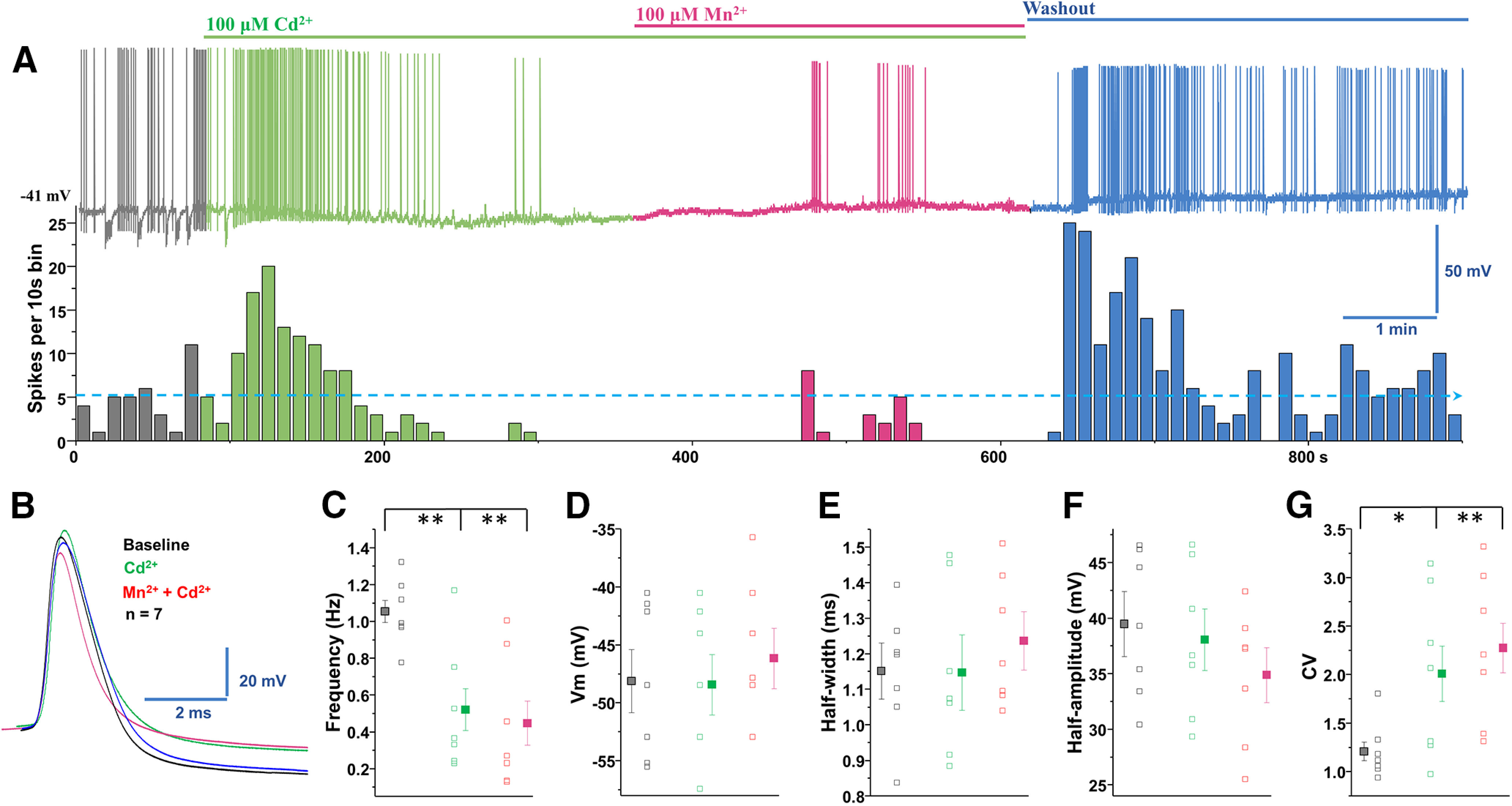
Cd2+ blockade of voltage-gated Ca2+ channels inhibited manganese stimulation of spontaneous firing activity of dopamine neurons. A, Top, Representative spontaneous spike activity of a dopamine neuron before and after exposure to Cd2+ (a nonselective voltage-gated Ca2+ channel blocker) and Cd2+ + Mn2+. Cd2+ blockade of voltage-gated Ca2+ channels inhibited Mn2+ stimulation of spontaneous firing activity. Bottom, Histogram of firing frequency from above trace showing that firing frequencies were lowered in Cd2+ only condition and following or Cd2+ + Mn2+ but return to baseline after washout. B, Superimposed representative single AP traces shown in A: baseline (black trace), 100 μm Cd2+ (green trace), 100 μm Mn2+ + Cd2+ (red trace), and washout (blue trace). C, Spontaneous firing rate significantly decreased following application of Cd2+ or Cd2+ + Mn2+ (F(2,18) = 8.3, p = 0.002, one-way ANOVA followed by Tukey's test, n = 7/group). D, Application of Cd2+ or Cd2+ + Mn2+ did not change the membrane potential. E, Half-width was measured at half-maximal voltage of AP; there was no change in the half-width of the Cd2+ and Cd2+ + Mn2+ experimental groups. F, Cd2+ or Cd2+ + Mn2+ treatments did not change the amplitude of AP. G, Both Cd2+ and Cd2+ + Mn2+ treatments increased the CVs of the interspike interval (F(2,18) = 4.6, p = 0.024, one-way ANOVA followed by Tukey's test, n = 7/group). *p < 0.05, **p < 0.01.