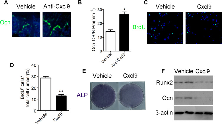
Figure 3.
Cxcl9 attenuates bone formation in vivo and in vitro. (A) Immunofluorescent staining of Ocn in the distal femur of OVX mice treated with vehicle or Cxcl9–neutralizing antibody (Anti-Cxcl9) with the first injection at the same day of OVX. Scale bar, 50 μm. (B) Number of osteocalcin positive osteoblasts (Ocn+ OB) on the bone surface was measured as cells per millimeter of perimeter in sections (/B.Pm). n=9 per group. (C) Representative confocal images of immunostaining of BrdU (green) in mouse primary calvarial osteoblasts. Scale bar, 100 μm. (D) Quantitative analysis of BrdU+ cells over total cells. n=9 per group. (E) ALP staining differentiated calvarial osteoblasts on the 7th day of osteogenic induction. (F) Western blot analysis of osteoblastic marker Runx2 and Ocn expression in differentiated calvarial osteoblasts on the 7th day. Data are shown as mean ± s.d. *P < 0.05, **P < 0.01 (Student’s t-test).

