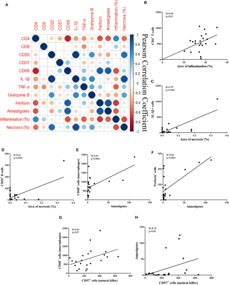
Figure 2.
Correlations between histopathological parameters and cellular markers in CL biopsies. (A) Clustered heatmap of Pearson correlation coefficients of the inflammatory cells, inflammation and necrosis; (B–H) Pearson correlation of the cells, areas of inflammation and necrosis, amastigotes and NK cells; (B) CD4+ T cells vs. inflammation; (C) IL-1β+ cells vs. necrosis; (D) CD20+ B cells vs. necrosis; (E) CD68+ macrophages vs. amastigotes; (F) perforin+ cells vs. amastigotes (G) CD68+ macrophages vs. CD57+ NK cells; (H) amastigotes vs. CD57+ NK cells.

