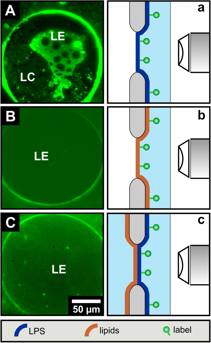
FIGURE 3.
LPS R45 free-standing planar monolayers lose their phase separation upon combination to an LPS R45/PL free-standing bilayer. Fluorescence microscopic images of (A) a LPS R45 monolayer, (B) a PL (PE:PG:CL; 81:17:2; w/w/w) monolayer, and (C) a LPS R45/PL planar bilayer reconstituted over an aperture. For fluorescence labeling of an individual membrane leaflet, the lipid monolayer was supplement with 1% NBD-PE which partitions into the liquid expanded (LE) domains. As with the solid-supported membranes, phase separation in free-standing lipid systems occurred only for the LPS R45 monolayer. Experiments were conducted in 100 mM KCl, 5 mM MgCl2, 5 mM HEPES at pH 7.0 and T = 37°C.

