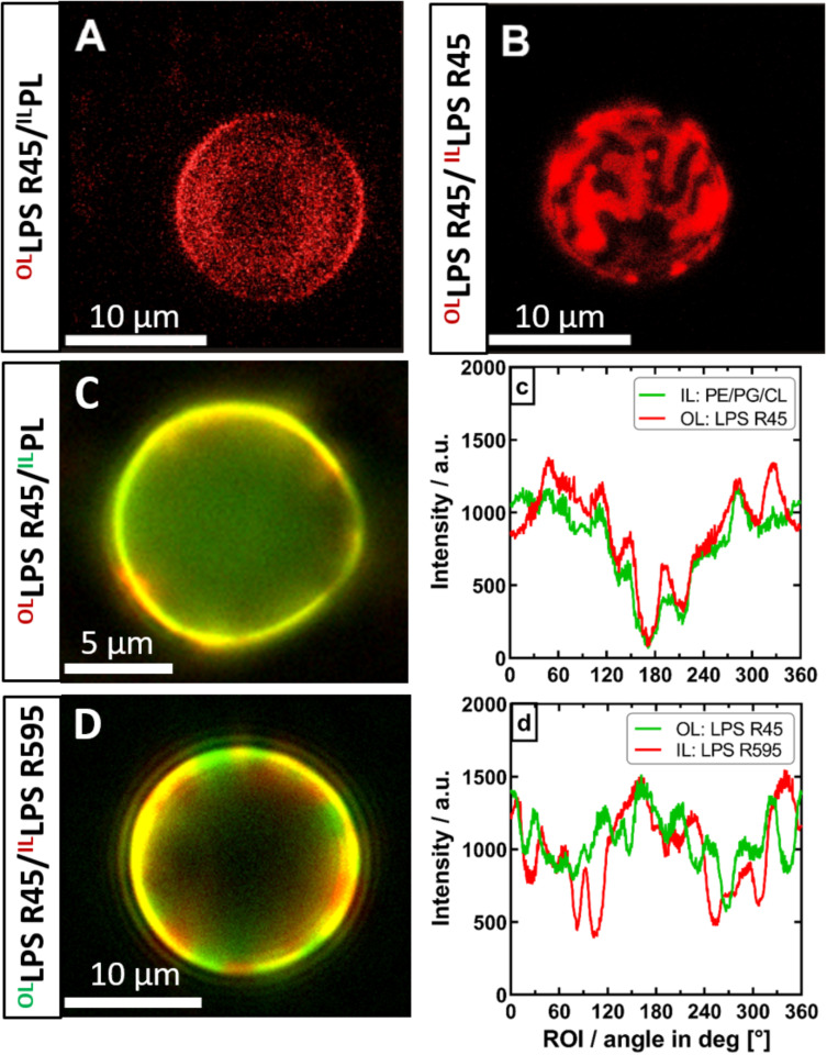
FIGURE 6.
The phase separation of the outer leaflet of asymmetric vesicles is depending on interactions with the inner leaflet. Both asymmetric and symmetric giant unilamellar vesicles were generated by the phase transfer method. (A) Asymmetric LPS R45/RhoPL vesicles expose no phase separation (PL refers to PE:PG:CL; 81:17:2; w/w/w). (B) Symmetric RhoLPS R45/RhoLPS R45 vesicles show dominant phase separation in the outer as well as in the inner leaflet (IL), exposing stripe and patch shaped domains. (C) Phase separation in asymmetric LPS R45/PL vesicles could not be enforced by lowering divalent cation content. (c) Fluorescence dye distribution analysis of IL and outer leaflet (OL). The fluorescent dye conjugates of both leaflets correlate and show an intensity minimum at approx. 180°. (D) Asymmetric FITCLPS R45/RhoLPS R595 vesicles show a dominant phase separation with areas wherein the fatty acid chains of the different LPS types match (yellow) and mismatch (red/green). (d) Fluorescence dye distribution analysis of the individual LPS leaflets giving areas of correlation and anti-correlation of the different fluorescence labels. All experiments were run out in 100 mM KCl buffer supplemented with either 5 mM MgCl2 and 5 mM HEPES (A+B,D) or 0.5 mM CaCl2x2H2O (C) at pH 7. Confocal laser scanning microscopy (A+B) was performed at 26°C. Images were taken with a Leica SP3. Z-series were performed and compiled for 3D visualization. Inverse fluorescence microscopy was performed at room temperature with an Olympus IX-81.

