Figure 3.
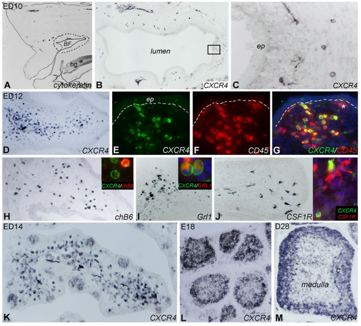
Localization of CXCR4 expression during bursal ontogeny. Immunocytochemistry of embryonic day (ED) 10 (A–C), ED12 (D–J), ED14 (K), ED18 (L), and 4 weeks old (ED28; M) chicken bursa of Fabricius. (A) The cytokeratin+ bursal epithelial anlage is a vesicle-like structure surrounded by the tail bud mesenchyme (dashed line). (B) A few round shaped CXCR4+ cells appear around the epithelial anlage. (C) Magnified image of the boxed area in (B). (D) Bursal folds are filled with CXCR4+ cells. (E–G) CD45 immunostaining confirms the hematopoietic origin of CXCR4+ cells whereas the ramified CD45+ cells do not express CXCR4 (* in G). chB6+ B cells (H) and Grl1+ granulocytes (I) are dispersed throughout the bursal fold and coexpress CXCR4 antigen (insets). (J) Ramified CSF1R+ cells are scattered throughout the bursa mesenchyme. Inset: double immunofluorescence staining shows that these cells do not express CXCR4. (K) ED14: CXCR4+ cells heavily infiltrate the bursa mesenchyme and colonize the follicle buds. (L) ED18: CXCR4+ cells are grouped in the lymphoid follicles. (M) After hatching only cortical B cells express CXCR4 antigen.

