Figure 4.
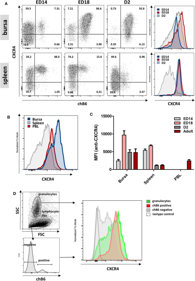
Varying CXCR4 surface expression during B cell ontogeny. (A) Lymphocytes were isolated from bursa and spleen at the indicated time points and analyzed for CXCR4 and chB6 expression by flow cytometry. Histograms were gated for chB6 positive cells. (B) CXCR4 expression on chB6 positive cells of a 4 weeks old bird. (C) MFI for anti-CXCR4 staining on chB6 positive cells. (D) Viable (7-AAD negative) cells from a ED14 bursa were separated into granulocytes and lymphocyte-like cells according to scatter properties; lymphocyte-like cells were further divided into chB6 positive and negative cells and CXCR4 expression was analyzed. (A,B,D) one representative of three independent experiments, (C) n = 3, mean ± SD.

