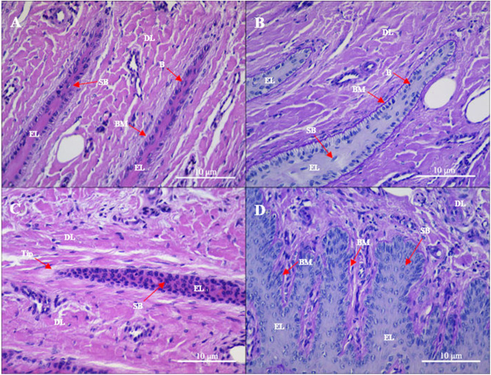
Figure 1.
Gross sections of the lamellae layer from (A,B) control and (C,D) oligofructose treated heifers. Labels in the sections: B, basal cells; SB, surprabasal cells; BM, basement membrane; DL, dermal lamellae; EL, epidermal lamellae; WBC, white blood cells; Tip, the tip of the epidermal lamellae). (A) H&E stain. The normal appearance of the dermo-epidermal junction consists of numerous interlocking lamellae. (B) Periodic acid–Schiff (PAS) stain. The tip of the epidermal lamellae is rounded and in a close association with the basement membrane. (C) H&E stain. Stretched epidermal lamellae and increased numbers of suprabasal cells are observed. (D) PAS stain. The basement membrane has an attenuated appearance; the basal cells are enlarged and wider in the dermal lamellae.

