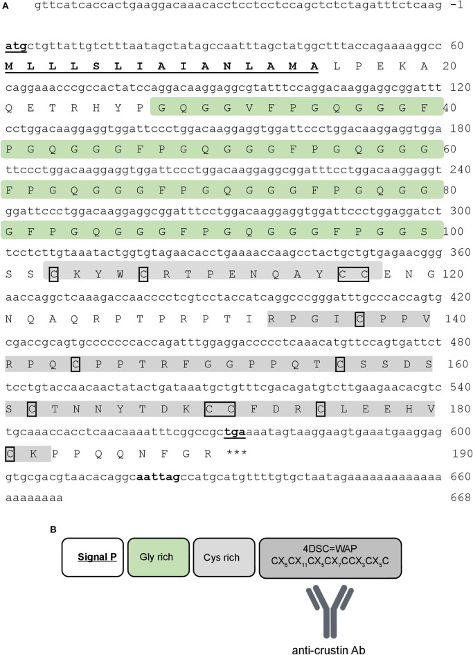
Figure 2.
Re-crustin sequence. (A) The full-length nucleotide (above) and predicted amino acid (below) sequences of Re-crustin cDNA from Rimicaris exoculata. The start and stop codons and the putative polyadenylation site are in bold and underlined. The signal peptide is underlined. The 12 conserved cysteine residues are framed. (B) The predicted organization of WAP domain is shown in the dark gray box.

