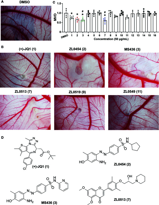
Figure 1.

Initial screening of select BET inhibitors to determine their effects on angiogenesis in the chick embryo CAM model. Representative inhibitory results on the growth of blood vessel branches by DMSO (A) or the selected BRD4 inhibitors (B), which showed strong positive inhibition (+++) of angiogenesis in the chick embryo CAM model. Each BET inhibitors (50 μg/ml) or DMSO was added directly onto the live 9-day-old chick embryo CAM model and incubated for 48 h. Then, the blood vessel network in the chicken embryo CAM was photographed by an OPTPRO 2007 image system. (C) The statistical results of microvessel density (MVD) analysis of the chick embryo CAM model treated with the selected BET inhibitors are shown in Table 1 . (D) Structures of (+)-JQ1 (1) (positive control), ZL0454 (2), MS436 (3), and ZL0513 (7). *P < 0.05 compared with the DMSO group, # P < 0.05 compared with the (+)-JQ1 (1) positive control group.
