
FIGURE 6.
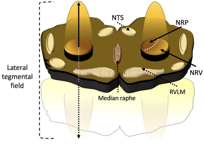
Artist rendition of a transverse slab of medulla the anatomic relationship between the medullary lateral tegmental field and rostral ventrolateral medulla. Propriobulbar interneuronal microcircuit oscillators constituting the medullary lateral tegmental field residing within the caudolateral medulla extend in line with rostromedial ascent toward the tip of the rostral ventrolateral medulla, thus presenting the architectonic appearance of the cometary tail of the rostral ventrolateral medulla in vivo. The medullary lateral tegmental field contains barosensitive units providing sympathoexcitatory and sympathoinhibitory propriobulbar inputs to the rostral ventrolateral medulla. The discharge of medullary lateral tegmental field neurons precedes unitary activity of rostral ventrolateral medullary units and exhibits correlation with bulbospinal presympathetic neuronal and cardiac sympathetic neural efferent discharge. The medullary lateral tegmental field contributes prominently to basal sympathoexcitation and reflexive changes in sympathetic tone, mediating baroreceptor, chemoreceptor, Bezold-Jarisch, somatoautonomic, and visceroautonomic reflexes via N-methyl-D-aspartate and non-N-methyl-D-aspartate-dependent mechanisms. NRP, nucleus reticularis paragigantocellularis; NRV, nucleus reticularis ventralis; NTS, nucleus tractus solitarius; RVLM, rostral ventrolateral medulla.