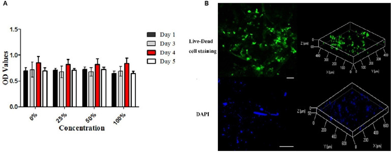
FIGURE 5.
Cytotoxicity of the decellularized meniscus ECM scaffold and recellularization in vitro. (A) Cytotoxicity assay of DM: the proliferative activities of the cells cultured in standard medium and those cultured in the extracts at different concentrations. (B) Fluorescence Live-Dead cell (Scale bar: 30 μm) and DAPI (Scale bar: 100 μm) staining images (two and three dimensions) demonstrating seeding of BMSCs on DM ECM scaffolds at 2 weeks.

