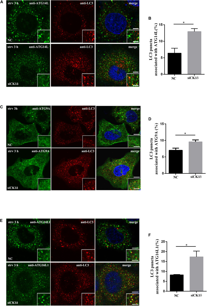
FIGURE 3.
Depletion of CK1δ results in increased association of LC3 and multiple ATG proteins. (A,B) Immunostaining of LC3 and ATG14L using endogenous antibodies. HeLa cells depleted of CK1δ and control HeLa cells were starved for 3 h. Scale bars, 5 μm. Scale bars in insets, 2 μm. Quantification of the percentage of LC3 puncta that associate with ATG14L puncta is shown in panel (B). Cells from three separate experiments (150 in total) were examined. Error bars represent SEM; *p < 0.05, Student’s t test. (C,D) Immunostaining of LC3 and ATG9A using endogenous antibodies. HeLa cells depleted of CK1δ and control HeLa cells were starved for 3 h. Scale bars, 5 μm. Scale bars in insets, 2 μm. Quantification of the percentage of LC3 puncta that associate with ATG9A puncta is shown in panel (D). Cells from three separate experiments (150 in total) were examined. Error bars represent SEM; *p < 0.05. Student’s t test. (E,F) Immunostaining of LC3 and ATG16L1 using endogenous antibodies. HeLa cells depleted of CK1δ and control HeLa cells were starved for 3 h. Scale bars, 5 μm. Scale bars in insets, 2 μm. Quantification of the percentage of LC3 puncta that associate with ATG16L1 puncta is shown in panel (D). Cells from three separate experiments (150 in total) were examined. Error bars represent SEM; *p < 0.05. Student’s t test.

