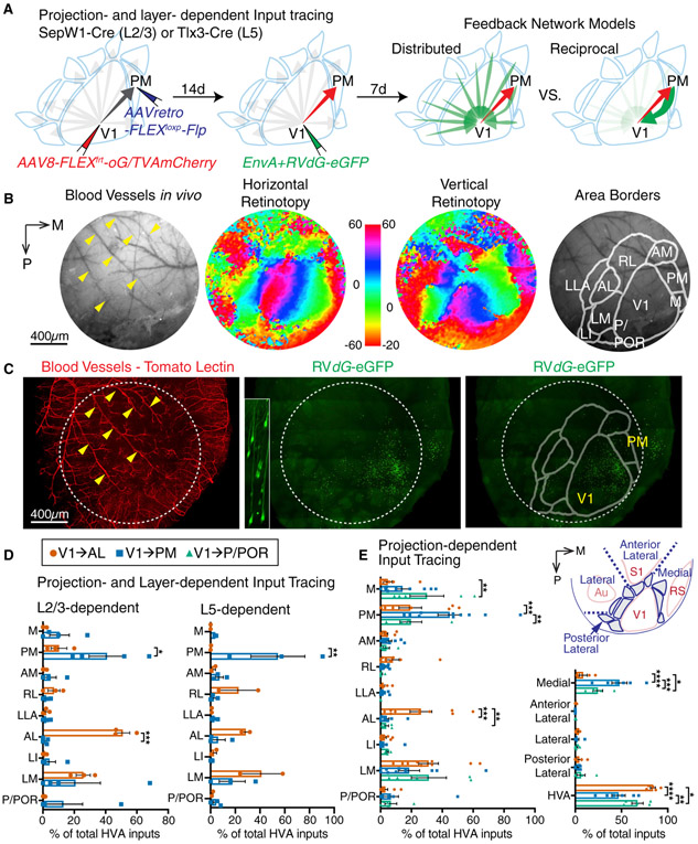
Figure 3. Projection- and Layer-dependent Input Tracing of V1→AL and V1→PM Neurons.
(A) Schematic showing the experimental design on projection- and layer-dependent monosynaptic input tracing by G-deleted rabies (left and middle). Two possible feedback connectivity models depending on projection (right). (B) Intrinsic signal imaging to define higher visual area borders. (C) Reconstruction of rabies-traced and blood vessel stained visual cortex using light sheet microscopy. (D) Summary of long-range feedback inputs onto V1→AL and V1→PM neurons in L2/3 (left) or L5 (right). (E) Summary of long-range feedback inputs onto V1→AL, V1→PM, and V1→P/POR neurons. Values are reported as mean ± SEM. Statistics were calculated from two-way ANOVAs with Sidak’s or Turkey’s post-hoc multiple-comparison tests for panels D and E, respectively. *p<0.05; **p< 0.01; ***p< 0.001; ****p<0.0001. A, anterior; AL, anterolateral; AM, anteromedial; Au, auditory cortex; LI, interomediolateral; LM, lateromedial; M, medial; P, posterior; PM, posteromedial; POR, postrhinal; RL, rostrolateral; RS, retrosplenial cortex; S1, primary somatosensory cortex; V1, primary visual cortex.

