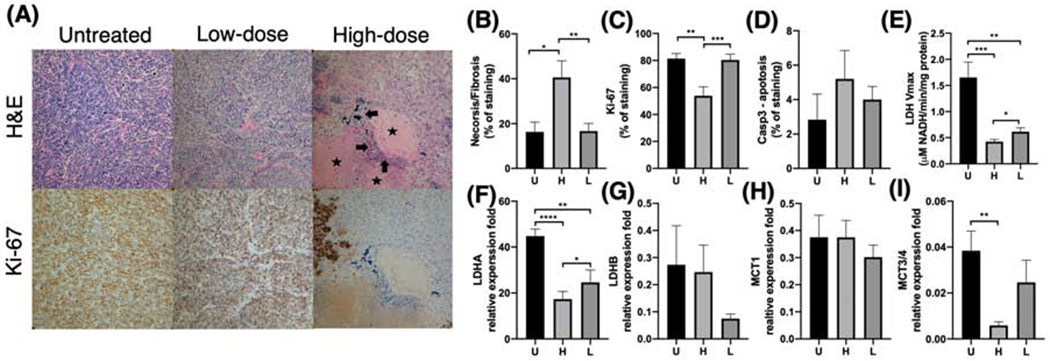
Figure 4:

Dose-dependent changes in immunohistochemical and molecular markers. Tumor regions treated with high radiation dose showed significantly more necrosis/fibrosis (A: arrows - frank necrosis; stars - frank fibrosis) on H&E staining (B) and lower Ki-67 (proliferation) staining (C) compared to their corresponding low-dose regions as well as a separate cohort of untreated TRAMP tumors (20x magnification). Moreover, high-dose tumor regions showed lower lactate dehydrogenase (LDH) activity (E), LDHA expression (F), and MCT3/4 expression (I) compared to low-dose regions, consistent with the decreased hyperpolarized 13C lactate signal observed in the high dose regions. Additionally, high-dose regions showed slightly higher anti-caspase-3 (an apoptosis marker) staining (D), higher LDHB expression (G), and lower MCT1 (H) expression compared to low-dose regions or untreated tumors, but these differences were not statistically significant. (U = untreated, H = high, L = low; *, P < .05; **, P < .01; ***, P < .001; ****, p < .001)
