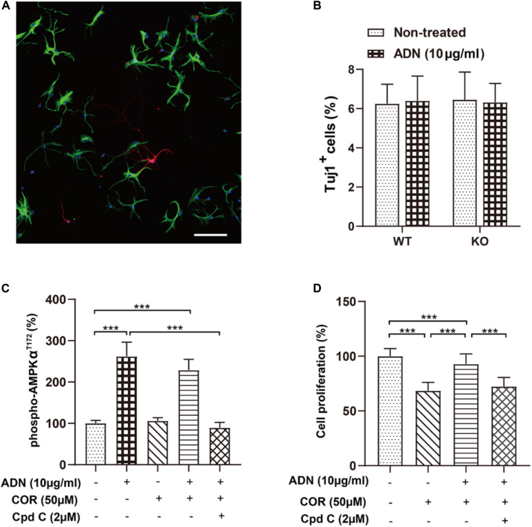
FIGURE 5.
Inhibition of the AMPK pathway diminishes adiponectin-enhanced cell proliferation in vitro. (A) The representative image showing that NPCs grown on coverslips after a 6-day culture in the induction medium differentiated into the Tuj1+ neuronal cells (red) and the GFAP+ glial cells (green) with the corresponding diverse morphologies. Scale bar: 100 μm. (B) Adding ADN into the medium to a final concentration of 10 μg/ml did not affect the percentage of induced neuronal cells in either WT or KO NPCs; the basal neuronal differentiation was unaltered after ADN KO. N = 6 coverslips/group from 3 independent experiments. (C) The phosphorylation of AMPK at the T172 site after application of ADN, COR and Cpd C. COR did not affect the level of phospho-AMPKT172, whereas Cpd C essentially abolished adiponectin-induced enhancement of AMPK phosphorylation. N = 4 independent experiments. (D) Cell proliferation following ADN, COR, and Cpd C treatments. Administration of COR significantly impaired neuronal proliferation which could be reversed by adiponectin addition, whereas inhibition of AMPK phosphorylation by Cpd C potently abolished the aforementioned proliferating effect of adiponectin. N = 3 independent experiments. Data are shown as means ± SD. ***p < 0.001 by one-way ANOVA and Tukey’s post hoc test. Tuj1, Neuronal Class III β-Tubulin (neuronal marker); GFAP, glial fibrillary acidic protein (glial marker); COR, corticosterone; ADN, adiponectin; Cpd C, Compound C (AMPK antagonist).

