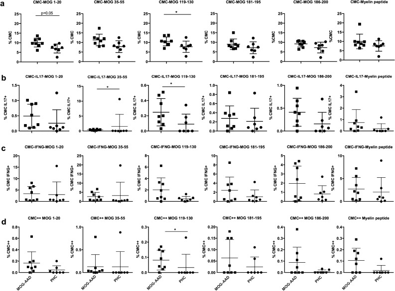
Figure 1.
Cytokine expression in MOG-reactive central memory cells (CMCs) from untreated MOG-AAD patients and pediatric healthy controls (PHCs): PBMCs were stimulated and cultured with 10 μg/ml of MOG peptides (p1-20, p35-55, p119-130, p181-195 and p186-200) and cocktail of myelin peptides (Myelin Phospholipid Protein 139–154 (PLP), Myelin Basic Protein (MBP) 13–32, MBP 111–129 and MBP 146–170) for 7 days followed by staining with antibodies and flow cytometry as described in Materials and Methods. Data was analyzed using Flowjo vX.0.7 and the graphs were made using GraphPadPrism version 8.4.2 (464) A Percentage of CMCs (CD4+CCR7+CD45RA−). B Percentage of IL17+producing CMCs (CD4+CCR7+CD45RA-, IL17+). C Percentage of IFNγ+producing CMCs (CD4+CCR7+CD45RA−,IFNγ +). D Percentage of IL17+ and IFNγ+ producing CMCs (CD4+CCR7+CD45RA−,IL17+,IFNγ+). MOG-AAD n = 8, PHC n = 7, Mann–Whitney test, *P < 0.05.

