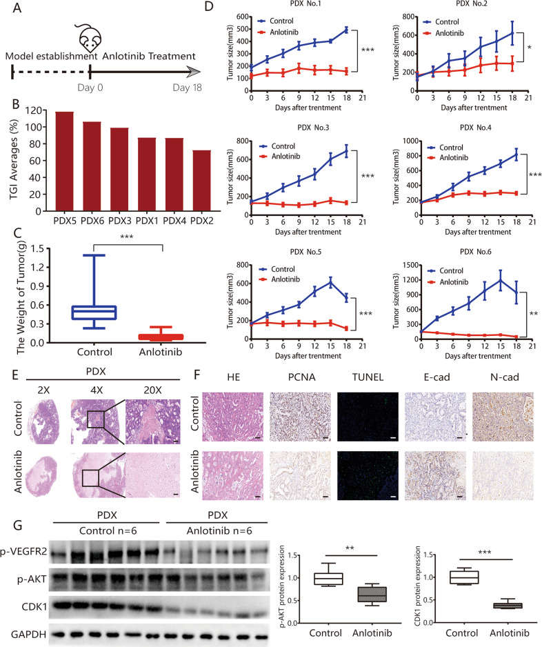
Fig. 3. Anlotinib exhibits effective antitumor activity with low drug toxicity in PDX models.
a Timeline of the experiments in vivo; b waterfall plot of anlotinib response after treatment in six PDX models; c there were significant differences in tumor weights between the anlotinib treatment group and the control group (0.58 ± 0.08 vs. 0.10 ± 0.01, respectively); d tumor growth curves for each PDX case; e representative hematoxylin and eosin staining of tumor tissues from PDX models, with or without anlotinib treatment; f anlotinib treatment group had substantial decreases in proliferation (PCNA) rates and increases in apoptosis (TUNEL) index numbers, compared with the control group. The expression of E-cadherin was significantly downregulated, while the expression of N-cadherin was upregulated, based on the immunohistochemistry (IHC) assay after anlotinib treatment; g expression of p-VEGFR2, p-AKT, CDK1 in PDX tumors was detected using western blot assay. *P < 0.05; **P < 0.01; ***P < 0.001. Scale bars = 100 μm.

