Figure 6.
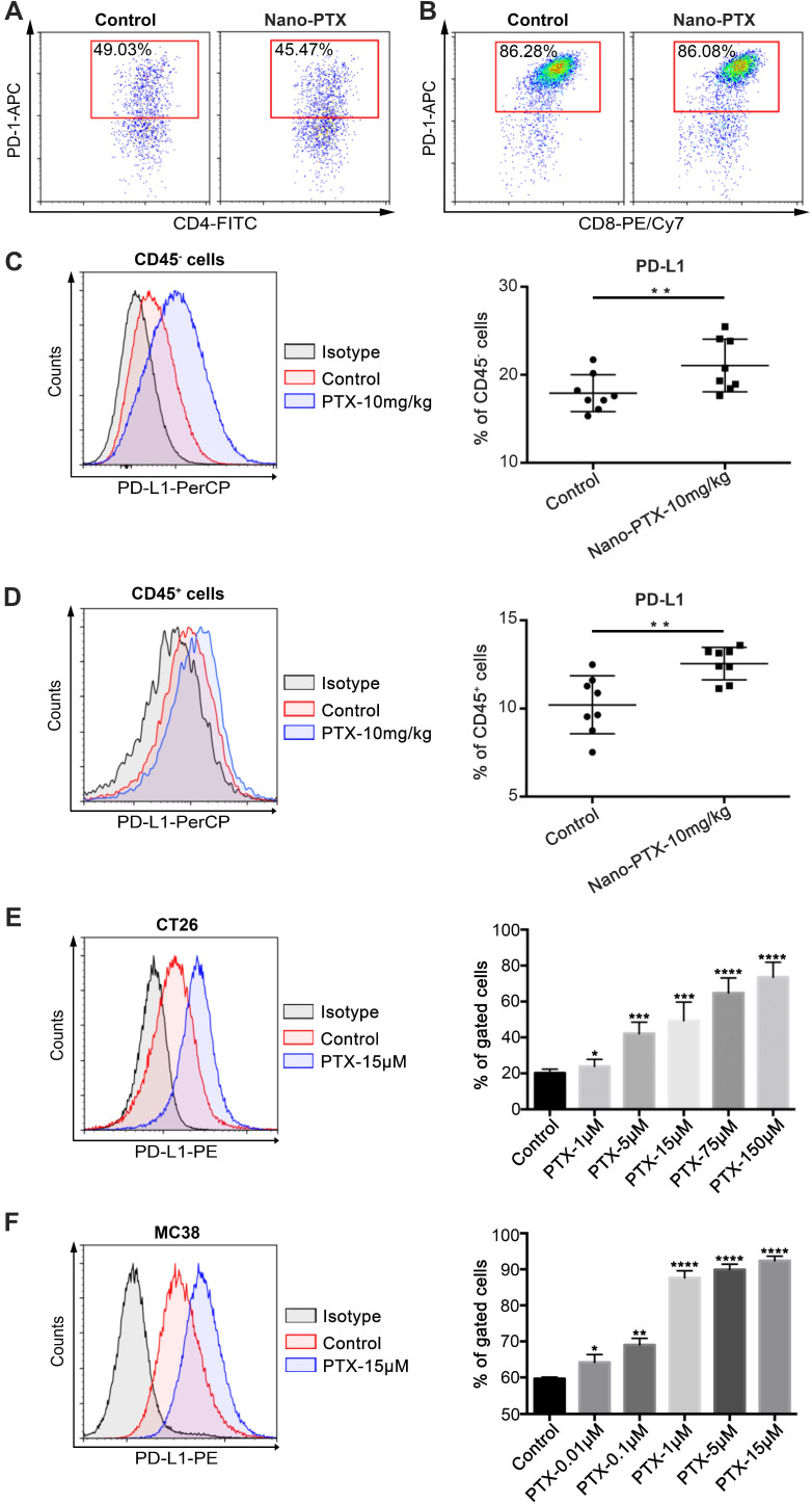
PTX treatment up-regulates PD-L1 expression within tumor microenvironment. A-D Mice with established CT26 tumors were treated with nano-PTX as described in Figure 5G, tumors were harvested and analyzed by flow cytometry on day 20. Representative flow data of PD-1 gated on CD4+ T cells (A) and CD8+ T cells (B) were shown. The percentages of PD-L1+ cells gated on CD45- cells (C) and CD45+ cells (D) were shown, n = 8 mice per group. E-F CT26 cells (E) and MC38 cells (F) were treated with PTX for 24 h in different concentrations as indicated. The PD-L1 expression was detected using flow cytometry, n = 8 replicates. Mean ±SEM was shown. * P < 0.05, ** P < 0.01, *** P < 0.001, **** P < 0.0001.

