Figure 8.
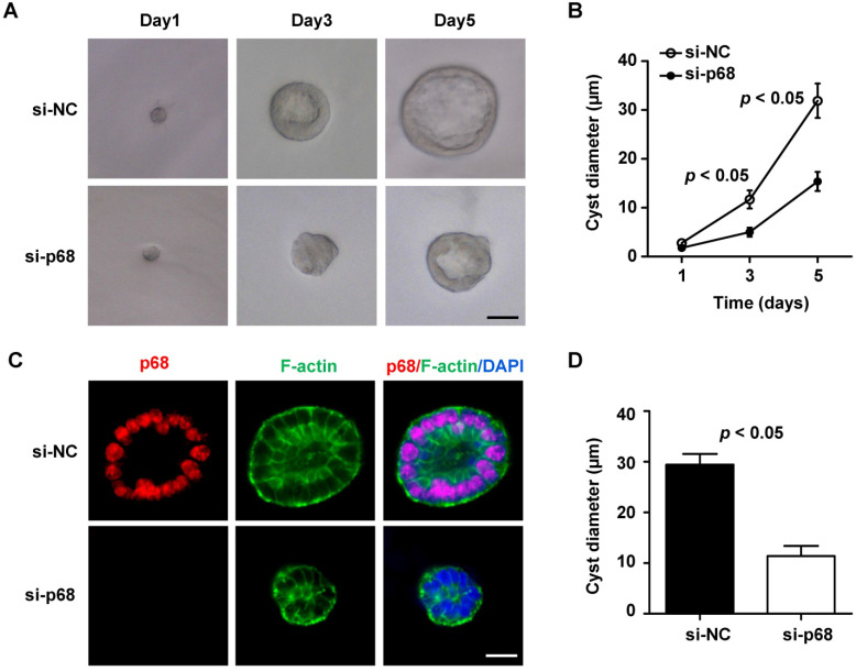
Knockdown of p68 delays spheroid (cyst) growth in mIMCD3 cell 3D cultures. (A) Light micrographs of cysts formed in Matrigel with mIMCD3 cells transfected NC-siRNA or p68-siRNA at the days 1, 3 and 5. Each series of photographs showed the same cyst on successive days in 3D cultures. Scale bars, 10 µm. (B) Cyst diameters of 3D cultures of mIMCD3 cells transfected with NC-siRNA or p68-siRNA on day 5. A total of 50 spheroids for each group from three different experiments were evaluated. (C) Spheroids were immunofluorescence-stained with antibodies against p68 and FITC-conjugated phalloidin for F-actin as well as DAPI. Spheroid morphology from mIMCD3 cells transfected with NC-siRNA or p68-siRNA (on day 5) judged by focusing to the equator plane of each spheroid using the z-axis of a confocal microscope (Leica). A total of 50 spheroids for each group from three different experiments were evaluated; scale bar, 10 µm. (D) The cyst diameters (µm) for NC-siRNA or p68 knockdown are plotted in a bar diagram. The area bounded by the F-actin of the cells is determined at the level of the largest lumen diameter of each cyst. Cysts that did not develop an open lumen were assigned an area of 0.

