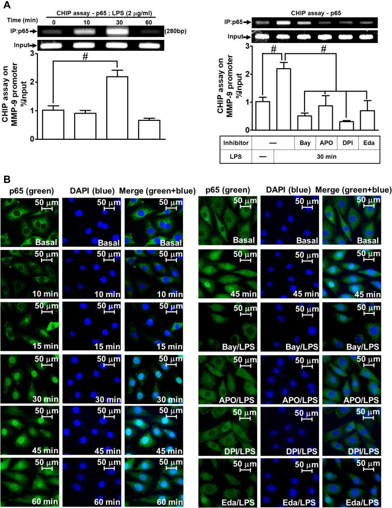
Figure 5.
NF-κB p65 nuclear translocation and binding with MMP-9 promoter element are involved in LPS-induced responses in RBA-1 cells. (A) Cells were incubated with LPS (2 μg/mL) for the indicated time intervals (0, 10, 30, and 60 min) (left panel). Cells were pretreated with or without APO (30 μM), DPI (10 μM), Edaravone (30 μM), or Bay11-7082 (1 μM) for 1 h and then incubated with LPS (2 μg/mL) for 30 min (right panel). The levels of NF-κB p65 binding to the MMP-9 promoter element were determined by a chromatin immunoprecipitation (ChIP) assay. (B) Cells were incubated with LPS (2 μg/mL) for the indicated time intervals (0, 10, 15, 30, 45, and 60 min). Cells were pretreated with or without APO (30 μM), DPI (10 μM), Edaravone (30 μM), or Bay11-7082 (1 μM) for 1 h and then incubated with LPS (2 μg/mL) for 45 min. The NF-κB p65 nuclear translocation was determined by immunofluorescence staining. The figure represents one of three individual experiments. Scale bar represents 50 µm. Data are expressed as mean ± SEM of three independent experiments. # p < 0.01 as compared with the cells exposed to vehicle or LPS, as indicated.

