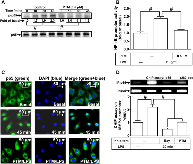
Figure 7.
Pristimerin attenuates LPS-induced NF-κB activity in RBA-1 cells. (A) Cells were pretreated with or without pristimerin (0.5 µM) for 1 h and then incubated with LPS (2 µg/mL) for the indicated time intervals (10, 30, and 45 min). The phosphorylation of NF-κB p65 was detected by Western blot. (B) Cells were pretreated with or without pristimerin (0.5 µM) and then incubated with LPS (2 μg/mL) for 4 h. NF-κB promoter luciferase activity was detected. (C) Cells were pretreated with or without pristimerin (0.5 µM) for 1 h, and then the LPS-triggered NF-κB p65 nuclear translocation was determined by immunofluorescence staining. The figure represents one of three individual experiments. Scale bar represents 50 µm. (D) Cells were pretreated with or without pristimerin (0.5 µM) or Bay11-7082 (1 μM) for 1 h, and then the levels of NF-κB p65 binding with MMP-9 promoter element region triggered by LPS were determined by a ChIP assay. Data are expressed as mean ± SEM of three independent experiments. # p < 0.01 as compared with the cells exposed to vehicle or LPS, as indicated.

