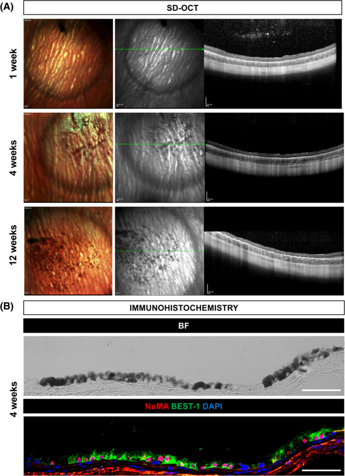
FIGURE 5.

Subretinal integration of hESC‐RPE in the albino rabbit eye. A, Multicolor‐confocal scanning laser ophthalmoscopy and SD‐OCT images of representative rabbits that received hESC‐RPE cells subretinally at 1, 4, and 12 weeks after transplantation. Green lines indicate the SD‐OCT scan plane. B, Representative BF and immunofluorescent images of NuMA and BEST‐1 staining of integrated hESC‐RPE in the rabbit subretinal space at 4 weeks after transplantation. Scale bars = 200 μm (A), 50 μm (B). BF, bright‐field; hESC, human embryonic stem cell; RPE, retinal pigment epithelial
