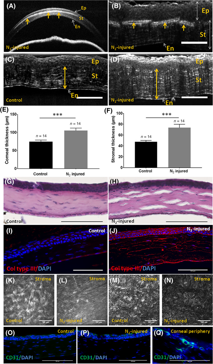
FIGURE 2.

Corneal stromal characterization 3 weeks after liquid nitrogen application. A,B, OCT and FFOCM cross sections of N2‐injured cornea, respectively. Arrows show the presence of an opaque zone in corneal stroma. C,D, FFOCM images used to determine corneal stromal and total corneal thicknesses with the ImageJ software before (C) and 3 weeks after N2 application (D). E,F, Corneal and stromal thicknesses were significantly higher (P = .0004) in injured vs control corneas 3 weeks after N2 application. G,H, Histological analysis with hematoxylin and eosin staining of control and N2‐injured corneas, respectively. I,J, Immunofluorescence analysis for collagen type III, in control vs N2‐injured corneas. K,L, IVCM images of normal (K) and N2‐injured (L) cornea showing the absence of blood vessel in corneal stroma. M,N, FFOCM en face images of normal (M) and N2‐injured (N) cornea showing the absence of blood vessel in corneal stroma. O‐Q, CD31 negative staining in normal (O) and N2‐injured (P) cornea. Positive CD31 staining was observed in corneal periphery (Q). Scale bars = 100 μm. Error bars = ±SEM. ***P < .001. FFOCM, full field optical coherence microscopy; IVCM, in vivo confocal microscopy; OCT, optical coherence tomography
