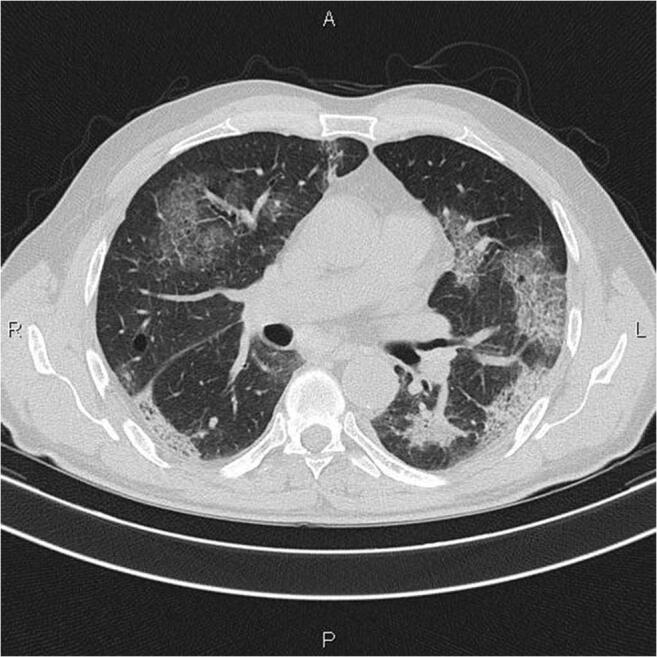
Fig. 2.

CT image from a confirmed COVID-19 case showing multiple patchy and light consolidations in both lungs and grid-like thickness of interlobular septae. Note: Radiograph image of confirmed COVID-19 case (36), courtesy of Dr. Mohammad Taghi Niknejad, Radiopaedia.org, rID: 75607 [21]
