
Figure 7.
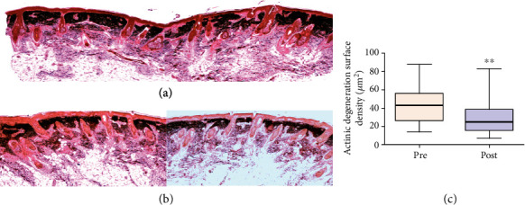
Photomicrographs representative of histological sections of biopsies before and after treatment with ADSCs. (a) Pretreatment biopsies showing intense actinic elastosis in the papillary and reticular dermis. (b) After treatment, a reduction in the elastotic mass in the dermis was observed. Stained: oxidized orcein. (c) Graph of the surface density of the reactive material for oxidized orcein in zone 2 reveals a significant reduction in elastosis (p = 0.005).