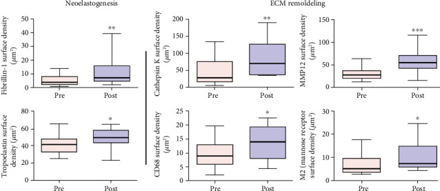
Figure 8.

Immunohistochemical analysis after ADSC skin therapy into two aspects: neoelastogenesis represented by anti-fibrillin immune labelling that showed overall increase of fibrillin, including in the zone 1 (p = 0.001), and anti-tropoelastin immune labelling that showed increase of the tropoelastin reactive material in the post treatment biopsy (p < 0.05). Extracellular matrix (ECM) remodeling was represented by anti-cathepsin K immunostaining that revealed an increase in quantity of CAT-K immune-labelled cells in the dermis of a posttreated skin biopsy (p = 0.011), anti-MMP12 immunostaining representation of percentage of MMP12 in the skin biopsies significantly increased after ADSC treatment (p = 0.005), anti-CD68 and CD206 (mannose receptor (MR)) macrophage immunostaining in sun-exposed facial skin before and after ADSC injection that showed a significant increase after ADSC treatment in the dermis of posttreated skin biopsy (p < 0.05).
