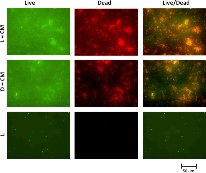
FIGURE 6.

Fluorescence microscopy images of live/dead staining of the PDI samples of B. subtilis when 1 mg/ml of the dye extract (CM) was used in water. CM, cold methanol dye extract. L + CM is the light experiment (dye extract with light illumination at 10 mW/cm2 for 20 min); D + CM is the dark control experiments (dye extract without light illumination for 20 min); and L is “light only” control experiment (no dye extracts but illuminated with light at 10 mW/cm2 for 20 min)
