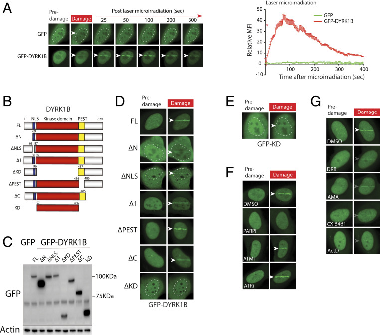
Fig. 3.
DYRK1B is recruited to laser-induced DSBs. (A) Cells expressing GFP-DYRK1B or GFP alone were laser microirradiated and time-lapse images were captured to analyze protein accumulation at DNA damage tracks. Quantification of GFP-DYRK1B or GFP accumulation at DSBs was performed. Arrowheads denote sites of laser microirradiation. (B and C) Schematics and steady-state expression level of DYRK1B and its deletion mutant are shown. FL, full length; KD, kinase domain; NLS, nuclear localization signal; PEST, proline-, glutamic acid-, serine-, and threonine-rich domain. (D and E) U2OS cells expressing GFP-DYRK1B or mutants (B) were laser microirradiated. Representative images from predamaged and laser-damaged (100 s postmicroirradiation) cells are shown. (F and G) U2OS cells expressing GFP-DYRK1B were pretreated with the indicated inhibitors prior to laser microirradiation. Cells were processed as in A and representative images are shown as in D.

