Fig. 3.
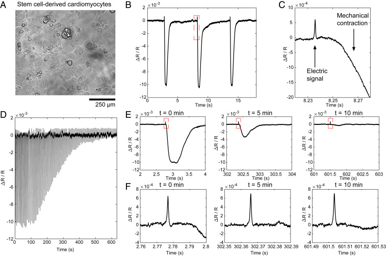
ECORE optical recording in stem cell–derived cardiomyocytes. (A) Bright-field image of stem cell–derived cardiomyocytes cultured in a PEDOT film. (B) Typical ECORE optical recording of cardiomyocytes. (C) The enlarged part of B shows the electrical signal as a small spike occurring about 15 ms before the onset of the cell’s mechanical contraction. (D) The ECORE recording shows a gradually decreased signal upon the application of 12.5 μM blebbistatin over 10 min. (E) Enlarged parts of D at 0, 5, and 10 min show a drastic decrease of the mechanical contraction. (F) Zoomed parts of E show that electric spikes remain the same at 0, 5, and 10 min.

